 Movie
Movie Controller


+ Open data

- Basic information
| Entry | Database: PDB / ID: 4y5w | ||||||
|---|---|---|---|---|---|---|---|
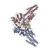
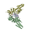
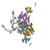
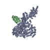
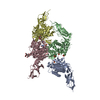
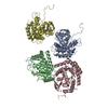
| Title | Transcription factor-DNA complex | ||||||
 Components Components |
| ||||||
 Keywords Keywords | TRANSCRIPTION/DNA / regulation / DNA binding / innate immune / TRANSCRIPTION-DNA complex | ||||||
| Function / homology |  Function and homology information Function and homology informationregulation of mast cell proliferation / mammary gland morphogenesis / positive regulation of isotype switching to IgE isotypes / negative regulation of type 2 immune response / T-helper 1 cell lineage commitment / STAT6-mediated induction of chemokines / interleukin-4-mediated signaling pathway / isotype switching to IgE isotypes / mammary gland epithelial cell proliferation / growth hormone receptor signaling pathway via JAK-STAT ...regulation of mast cell proliferation / mammary gland morphogenesis / positive regulation of isotype switching to IgE isotypes / negative regulation of type 2 immune response / T-helper 1 cell lineage commitment / STAT6-mediated induction of chemokines / interleukin-4-mediated signaling pathway / isotype switching to IgE isotypes / mammary gland epithelial cell proliferation / growth hormone receptor signaling pathway via JAK-STAT / sperm head-tail coupling apparatus / cell surface receptor signaling pathway via JAK-STAT / Downstream signal transduction / RNA polymerase II transcription regulatory region sequence-specific DNA binding / defense response / response to peptide hormone / RNA polymerase II transcription regulator complex / transcription coactivator binding / cytokine-mediated signaling pathway / regulation of cell population proliferation / positive regulation of cold-induced thermogenesis / Interleukin-4 and Interleukin-13 signaling / DNA-binding transcription activator activity, RNA polymerase II-specific / protein phosphatase binding / DNA-binding transcription factor activity, RNA polymerase II-specific / RNA polymerase II cis-regulatory region sequence-specific DNA binding / DNA-binding transcription factor activity / regulation of transcription by RNA polymerase II / chromatin / negative regulation of transcription by RNA polymerase II / positive regulation of transcription by RNA polymerase II / nucleoplasm / identical protein binding / nucleus / cytoplasm / cytosol Similarity search - Function | ||||||
| Biological species |  Homo sapiens (human) Homo sapiens (human) | ||||||
| Method |  X-RAY DIFFRACTION / SYNCHROTRON / MOLECULAR REPLACEMENT / Resolution: 3.104 Å X-RAY DIFFRACTION / SYNCHROTRON / MOLECULAR REPLACEMENT / Resolution: 3.104 Å | ||||||
 Authors Authors | Li, J. / Niu, F. / Ouyang, S. / Liu, Z. | ||||||
 Citation Citation | Journal: Proc.Natl.Acad.Sci.USA / Year: 2016 Title: Structural basis for DNA recognition by STAT6 Authors: Li, J. / Rodriguez, J.P. / Niu, F. / Pu, M. / Wang, J. / Hung, L.W. / Shao, Q. / Zhu, Y. / Ding, W. / Liu, Y. / Da, Y. / Yao, Z. / Yang, J. / Zhao, Y. / Wei, G.H. / Cheng, G. / Liu, Z.J. / Ouyang, S. | ||||||
| History |
|
- Structure visualization
| Structure viewer | Molecule: MolmilJmol/JSmol |
|---|
- Downloads & links
-Download
| PDBx/mmCIF format | 4y5w.cif.gz | 434.4 KB | Display | PDBx/mmCIF format |
|---|---|---|---|---|
| PDB format | pdb4y5w.ent.gz | 348.1 KB | Display | PDB format |
| PDBx/mmJSON format | 4y5w.json.gz | Tree view | PDBx/mmJSON format | |
| Others |  Other downloads Other downloads |
-Validation report
| Arichive directory | https://data.pdbj.org/pub/pdb/validation_reports/y5/4y5wftp://data.pdbj.org/pub/pdb/validation_reports/y5/4y5w | HTTPS FTP |
|---|
-Related structure data
| Related structure data |  4y5uSC 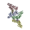 5d39C S: Starting model for refinement C: citing same article ( |
|---|---|
| Similar structure data |


-Links
 PDBj
PDBj


- Assembly
| Deposited unit | 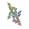
| ||||||||
|---|---|---|---|---|---|---|---|---|---|
| 1 | 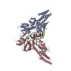
| ||||||||
| 2 | 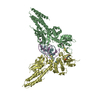
| ||||||||
| Unit cell |
|
-Components
| #1: Protein | Mass: 61577.602 Da / Num. of mol.: 4 / Fragment: UNP residues 113-658 Source method: isolated from a genetically manipulated source Source: (gene. exp.) Homo sapiens (human) / Gene: STAT6 / Plasmid: pMCSG7 / Production host:  #2: DNA chain | Mass: 6824.442 Da / Num. of mol.: 2 / Source method: obtained synthetically / Source: (synth.) Homo sapiens (human)#3: DNA chain | Mass: 6677.328 Da / Num. of mol.: 2 / Source method: obtained synthetically / Source: (synth.) Homo sapiens (human)#4: Water | ChemComp-HOH / |  Mass: 18.015 Da / Num. of mol.: 231 / Source method: isolated from a natural source / Formula: H2O Mass: 18.015 Da / Num. of mol.: 231 / Source method: isolated from a natural source / Formula: H2OHas protein modification | Y | |
|---|
-Experimental details
-Experiment
| Experiment | Method: X-RAY DIFFRACTION |
|---|
- Sample preparation
| Crystal | Density Matthews: 3.17 Å3/Da / Density % sol: 62.65 % Description: THE ENTRY CONTAINS FRIEDEL PAIRS IN F_PLUS/MINUS COLUMNS. |
|---|---|
| Crystal grow | Temperature: 289 K / Method: vapor diffusion, hanging drop Details: 0.1M citrate (pH 5.6), 0.1M NaCl, 20% isopropyl alcohol and 8% PEG4000, VAPOR DIFFUSION, HANGING DROP, temperature 289K |
-Data collection
| Diffraction | Mean temperature: 100 K |
|---|---|
| Diffraction source | Source: SYNCHROTRON / Site: SSRF  / Beamline: BL19U1 / Wavelength: 0.98 Å / Beamline: BL19U1 / Wavelength: 0.98 Å |
| Detector | Type: PSI PILATUS 6M / Detector: PIXEL / Date: Feb 6, 2014 |
| Radiation | Protocol: SINGLE WAVELENGTH / Monochromatic (M) / Laue (L): M / Scattering type: x-ray |
| Radiation wavelength | Wavelength: 0.98 Å / Relative weight: 1 |
| Reflection | Resolution: 3.1→50 Å / Num. obs: 57478 / % possible obs: 98 % / Redundancy: 3.5 % / Rsym value: 0.11 / Net I/σ(I): 11.2 |
| Reflection shell | Resolution: 3.1→3.21 Å / Redundancy: 3.4 % / Mean I/σ(I) obs: 1.65 / Rsym value: 0.4 / % possible all: 95.2 |
- Processing
| Software |
| ||||||||||||||||||||||||||||||||||||||||||||||||||||||||||||||||||||||||||||||||||||||||||||||||||||||||||||||||||||||||||||||||||||||||||||||||||||||||||
|---|---|---|---|---|---|---|---|---|---|---|---|---|---|---|---|---|---|---|---|---|---|---|---|---|---|---|---|---|---|---|---|---|---|---|---|---|---|---|---|---|---|---|---|---|---|---|---|---|---|---|---|---|---|---|---|---|---|---|---|---|---|---|---|---|---|---|---|---|---|---|---|---|---|---|---|---|---|---|---|---|---|---|---|---|---|---|---|---|---|---|---|---|---|---|---|---|---|---|---|---|---|---|---|---|---|---|---|---|---|---|---|---|---|---|---|---|---|---|---|---|---|---|---|---|---|---|---|---|---|---|---|---|---|---|---|---|---|---|---|---|---|---|---|---|---|---|---|---|---|---|---|---|---|---|---|
| Refinement | Method to determine structure: MOLECULAR REPLACEMENT Starting model: 4Y5U Resolution: 3.104→38.807 Å / SU ML: 0.43 / Cross valid method: FREE R-VALUE / σ(F): 1.97 / Phase error: 30.62 / Stereochemistry target values: ML
| ||||||||||||||||||||||||||||||||||||||||||||||||||||||||||||||||||||||||||||||||||||||||||||||||||||||||||||||||||||||||||||||||||||||||||||||||||||||||||
| Solvent computation | Shrinkage radii: 0.9 Å / VDW probe radii: 1.11 Å / Solvent model: FLAT BULK SOLVENT MODEL | ||||||||||||||||||||||||||||||||||||||||||||||||||||||||||||||||||||||||||||||||||||||||||||||||||||||||||||||||||||||||||||||||||||||||||||||||||||||||||
| Refinement step | Cycle: LAST / Resolution: 3.104→38.807 Å
| ||||||||||||||||||||||||||||||||||||||||||||||||||||||||||||||||||||||||||||||||||||||||||||||||||||||||||||||||||||||||||||||||||||||||||||||||||||||||||
| Refine LS restraints |
| ||||||||||||||||||||||||||||||||||||||||||||||||||||||||||||||||||||||||||||||||||||||||||||||||||||||||||||||||||||||||||||||||||||||||||||||||||||||||||
| LS refinement shell |
|
