 Movie
Movie Controller


+ Open data

- Basic information
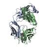
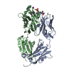
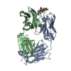
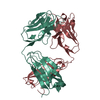
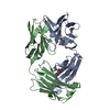
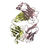
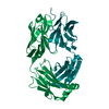
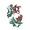
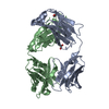
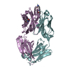
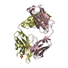
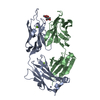
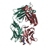
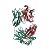
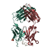
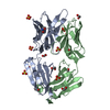
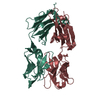
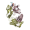
| Entry | Database: PDB / ID: 6ldx | ||||||
|---|---|---|---|---|---|---|---|
| Title | Structure antibody E6 in complex with methylated peptide | ||||||
 Components Components |
| ||||||
 Keywords Keywords | IMMUNE SYSTEM / methylation / antibody / phage display / biomolecular recognition | ||||||
| Function / homology |  Function and homology information Function and homology informationmitogen-activated protein kinase kinase kinase / MAP kinase kinase kinase activity / cellular response to mechanical stimulus / protein kinase activity / intracellular signal transduction / protein serine kinase activity / protein serine/threonine kinase activity / protein kinase binding / nucleoplasm / ATP binding ...mitogen-activated protein kinase kinase kinase / MAP kinase kinase kinase activity / cellular response to mechanical stimulus / protein kinase activity / intracellular signal transduction / protein serine kinase activity / protein serine/threonine kinase activity / protein kinase binding / nucleoplasm / ATP binding / metal ion binding / cytosol / cytoplasm Similarity search - Function | ||||||
| Biological species |   Homo sapiens (human) Homo sapiens (human) | ||||||
| Method |  X-RAY DIFFRACTION / SYNCHROTRON / MOLECULAR REPLACEMENT / Resolution: 1.8 Å X-RAY DIFFRACTION / SYNCHROTRON / MOLECULAR REPLACEMENT / Resolution: 1.8 Å | ||||||
 Authors Authors | Caaveiro, J.M.M. / Tsumoto, K. | ||||||
 Citation Citation | Journal: J.Biol.Chem. / Year: 2020 Title: Structural basis for antigen recognition by methylated lysine-specific antibodies. Authors: Ishii, M. / Nakakido, M. / Caaveiro, J.M.M. / Kuroda, D. / Okumura, C.J. / Maruyama, T. / Entzminger, K. / Tsumoto, K. | ||||||
| History |
|
- Structure visualization
| Structure viewer | Molecule: MolmilJmol/JSmol |
|---|
- Downloads & links
-Download
| PDBx/mmCIF format | 6ldx.cif.gz | 182.7 KB | Display | PDBx/mmCIF format |
|---|---|---|---|---|
| PDB format | pdb6ldx.ent.gz | 141.6 KB | Display | PDB format |
| PDBx/mmJSON format | 6ldx.json.gz | Tree view | PDBx/mmJSON format | |
| Others |  Other downloads Other downloads |
-Validation report
| Arichive directory | https://data.pdbj.org/pub/pdb/validation_reports/ld/6ldxftp://data.pdbj.org/pub/pdb/validation_reports/ld/6ldx | HTTPS FTP |
|---|
-Related structure data
| Related structure data | 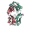 6ldvSC 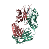 6ldwC 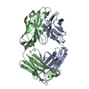 6ldyC S: Starting model for refinement C: citing same article ( |
|---|---|
| Similar structure data |


-Links
 PDBj
PDBj


- Assembly
| Deposited unit | 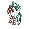
| ||||||||
|---|---|---|---|---|---|---|---|---|---|
| 1 |
| ||||||||
| Unit cell |
|
-Components
| #1: Antibody | Mass: 25795.686 Da / Num. of mol.: 1 Source method: isolated from a genetically manipulated source Source: (gene. exp.) Homo sapiens (human) |
|---|---|
| #2: Antibody | Mass: 25361.797 Da / Num. of mol.: 1 Source method: isolated from a genetically manipulated source Source: (gene. exp.) Homo sapiens (human) |
| #3: Protein/peptide | Mass: 1598.839 Da / Num. of mol.: 1 / Source method: obtained synthetically / Source: (synth.) Homo sapiens (human) / References: UniProt: Q9Y2U5*PLUS |
| #4: Chemical | ChemComp-CL /   Mass: 35.453 Da / Num. of mol.: 1 / Source method: obtained synthetically / Formula: Cl Mass: 35.453 Da / Num. of mol.: 1 / Source method: obtained synthetically / Formula: Cl |
| #5: Water | ChemComp-HOH /  Mass: 18.015 Da / Num. of mol.: 330 / Source method: isolated from a natural source / Formula: H2O Mass: 18.015 Da / Num. of mol.: 330 / Source method: isolated from a natural source / Formula: H2O |
| Has ligand of interest | Y |
-Experimental details
-Experiment
| Experiment | Method: X-RAY DIFFRACTION / Number of used crystals: 1 |
|---|
- Sample preparation
| Crystal | Density Matthews: 1.96 Å3/Da / Density % sol: 37.38 % |
|---|---|
| Crystal grow | Temperature: 293.15 K / Method: vapor diffusion, hanging drop / Details: 0.1 M potassium chloride 24% PEG3350 |
-Data collection
| Diffraction | Mean temperature: 100 K / Serial crystal experiment: N |
|---|---|
| Diffraction source | Source: SYNCHROTRON / Site: Photon Factory  / Beamline: BL-1A / Wavelength: 1 Å / Beamline: BL-1A / Wavelength: 1 Å |
| Detector | Type: DECTRIS EIGER X 4M / Detector: PIXEL / Date: Mar 16, 2019 |
| Radiation | Protocol: SINGLE WAVELENGTH / Monochromatic (M) / Laue (L): M / Scattering type: x-ray |
| Radiation wavelength | Wavelength: 1 Å / Relative weight: 1 |
| Reflection | Resolution: 1.8→67.94 Å / Num. obs: 39032 / % possible obs: 99.9 % / Redundancy: 23.2 % / CC1/2: 0.999 / Rmerge(I) obs: 0.092 / Rpim(I) all: 0.019 / Net I/σ(I): 23.3 |
| Reflection shell | Resolution: 1.8→1.9 Å / Redundancy: 13.5 % / Rmerge(I) obs: 0.468 / Mean I/σ(I) obs: 5.6 / Num. unique obs: 5534 / CC1/2: 0.875 / Rpim(I) all: 0.131 / % possible all: 99.1 |
- Processing
| Software |
| ||||||||||||||||||||||||||||||||||||||||||||||||||||||||||||||||||||||||||||||||||||||||||||||||||||
|---|---|---|---|---|---|---|---|---|---|---|---|---|---|---|---|---|---|---|---|---|---|---|---|---|---|---|---|---|---|---|---|---|---|---|---|---|---|---|---|---|---|---|---|---|---|---|---|---|---|---|---|---|---|---|---|---|---|---|---|---|---|---|---|---|---|---|---|---|---|---|---|---|---|---|---|---|---|---|---|---|---|---|---|---|---|---|---|---|---|---|---|---|---|---|---|---|---|---|---|---|---|
| Refinement | Method to determine structure: MOLECULAR REPLACEMENT Starting model: 6LDV Resolution: 1.8→67.94 Å / Cor.coef. Fo:Fc: 0.962 / Cor.coef. Fo:Fc free: 0.939 / SU B: 4.958 / SU ML: 0.08 / Cross valid method: THROUGHOUT / σ(F): 0 / ESU R: 0.122 / ESU R Free: 0.123 Details: HYDROGENS HAVE BEEN ADDED IN THE RIDING POSITIONS U VALUES : WITH TLS ADDED SF FILE CONTAINS FRIEDEL PAIRS UNDER I/F_MINUS AND I/F_PLUS COLUMNS.
| ||||||||||||||||||||||||||||||||||||||||||||||||||||||||||||||||||||||||||||||||||||||||||||||||||||
| Solvent computation | Ion probe radii: 0.8 Å / Shrinkage radii: 0.8 Å / VDW probe radii: 1.2 Å | ||||||||||||||||||||||||||||||||||||||||||||||||||||||||||||||||||||||||||||||||||||||||||||||||||||
| Displacement parameters | Biso max: 66.3 Å2 / Biso mean: 21.935 Å2 / Biso min: 13.75 Å2
| ||||||||||||||||||||||||||||||||||||||||||||||||||||||||||||||||||||||||||||||||||||||||||||||||||||
| Refinement step | Cycle: final / Resolution: 1.8→67.94 Å
| ||||||||||||||||||||||||||||||||||||||||||||||||||||||||||||||||||||||||||||||||||||||||||||||||||||
| Refine LS restraints |
| ||||||||||||||||||||||||||||||||||||||||||||||||||||||||||||||||||||||||||||||||||||||||||||||||||||
| LS refinement shell | Resolution: 1.8→1.847 Å / Rfactor Rfree error: 0 / Total num. of bins used: 20
| ||||||||||||||||||||||||||||||||||||||||||||||||||||||||||||||||||||||||||||||||||||||||||||||||||||
| Refinement TLS params. | Method: refined / Refine-ID: X-RAY DIFFRACTION
| ||||||||||||||||||||||||||||||||||||||||||||||||||||||||||||||||||||||||||||||||||||||||||||||||||||
| Refinement TLS group |
|
