 Movie
Movie Controller


+ Open data

- Basic information
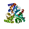
| Entry | Database: PDB / ID: 6lca | ||||||
|---|---|---|---|---|---|---|---|
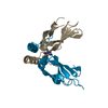
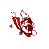
| Title | Crystal structure of human Dishevelled1 PDZ domain homotrimer | ||||||
 Components Components | Segment polarity protein dishevelled homolog DVL-1 | ||||||
 Keywords Keywords | SIGNALING PROTEIN / Wnt signaling pathway / Developmental protein / PROTEIN BINDING / Dvl | ||||||
| Function / homology |  Function and homology information Function and homology informationpositive regulation of protein localization to presynapse / Negative regulation of TCF-dependent signaling by DVL-interacting proteins / positive regulation of neuron projection arborization / presynapse assembly / non-canonical Wnt signaling pathway / WNT5:FZD7-mediated leishmania damping / dendritic spine morphogenesis / frizzled binding / PCP/CE pathway / Wnt signalosome ...positive regulation of protein localization to presynapse / Negative regulation of TCF-dependent signaling by DVL-interacting proteins / positive regulation of neuron projection arborization / presynapse assembly / non-canonical Wnt signaling pathway / WNT5:FZD7-mediated leishmania damping / dendritic spine morphogenesis / frizzled binding / PCP/CE pathway / Wnt signalosome / neurotransmitter secretion / clathrin-coated vesicle / regulation of postsynapse organization / WNT mediated activation of DVL / neural tube development / Disassembly of the destruction complex and recruitment of AXIN to the membrane / Wnt signaling pathway, planar cell polarity pathway / dendrite morphogenesis / heart looping / regulation of synaptic vesicle exocytosis / neuromuscular junction development / receptor clustering / outflow tract morphogenesis / lateral plasma membrane / canonical Wnt signaling pathway / protein localization to nucleus / neuronal dense core vesicle / positive regulation of excitatory postsynaptic potential / TCF dependent signaling in response to WNT / RHO GTPases Activate Formins / Degradation of DVL / synapse organization / beta-catenin binding / Schaffer collateral - CA1 synapse / small GTPase binding / regulation of protein localization / positive regulation of proteasomal ubiquitin-dependent protein catabolic process / growth cone / presynapse / cytoplasmic vesicle / dendritic spine / microtubule / neuron projection / postsynaptic density / protein stabilization / intracellular signal transduction / neuronal cell body / regulation of DNA-templated transcription / synapse / protein kinase binding / glutamatergic synapse / enzyme binding / positive regulation of transcription by RNA polymerase II / identical protein binding / cytosol Similarity search - Function | ||||||
| Biological species |  Homo sapiens (human) Homo sapiens (human) | ||||||
| Method |  X-RAY DIFFRACTION / SYNCHROTRON / MOLECULAR REPLACEMENT / Resolution: 2.4 Å X-RAY DIFFRACTION / SYNCHROTRON / MOLECULAR REPLACEMENT / Resolution: 2.4 Å | ||||||
 Authors Authors | Yasukochi, S. / Numoto, N. / Tenno, N. / Tenno, T. / Ito, N. / Hiroaki, H. | ||||||
 Citation Citation | Journal: To Be Published Title: Crystal structure of human Dishevelled1 PDZ domain homotrimer Authors: Yasukochi, S. / Numoto, N. / Tenno, N. / Tenno, T. / Ito, N. / Hiroaki, H. | ||||||
| History |
|
- Structure visualization
| Structure viewer | Molecule: MolmilJmol/JSmol |
|---|
- Downloads & links
-Download
| PDBx/mmCIF format | 6lca.cif.gz | 159 KB | Display | PDBx/mmCIF format |
|---|---|---|---|---|
| PDB format | pdb6lca.ent.gz | 115.5 KB | Display | PDB format |
| PDBx/mmJSON format | 6lca.json.gz | Tree view | PDBx/mmJSON format | |
| Others |  Other downloads Other downloads |
-Validation report
| Arichive directory | https://data.pdbj.org/pub/pdb/validation_reports/lc/6lcaftp://data.pdbj.org/pub/pdb/validation_reports/lc/6lca | HTTPS FTP |
|---|
-Related structure data
| Related structure data | 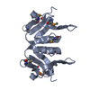 3fy5S S: Starting model for refinement |
|---|---|
| Similar structure data |


-Links
 PDBj
PDBj


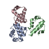
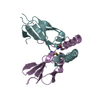
- Assembly
| Deposited unit | 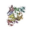
| ||||||||||||
|---|---|---|---|---|---|---|---|---|---|---|---|---|---|
| 1 | 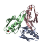
| ||||||||||||
| 2 | 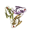
| ||||||||||||
| 3 | 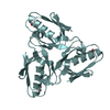
| ||||||||||||
| 4 | 
| ||||||||||||
| Unit cell |
|
-Components
| #1: Protein | Mass: 10771.055 Da / Num. of mol.: 8 / Mutation: C338A,W339T Source method: isolated from a genetically manipulated source Source: (gene. exp.) Homo sapiens (human) / Gene: DVL1 / Plasmid: pGEX-6P3 / Production host:  #2: Chemical | ChemComp-SO4 /   Mass: 96.063 Da / Num. of mol.: 8 / Source method: obtained synthetically / Formula: SO4 Mass: 96.063 Da / Num. of mol.: 8 / Source method: obtained synthetically / Formula: SO4#3: Water | ChemComp-HOH / |  Mass: 18.015 Da / Num. of mol.: 22 / Source method: isolated from a natural source / Formula: H2O Mass: 18.015 Da / Num. of mol.: 22 / Source method: isolated from a natural source / Formula: H2OHas ligand of interest | N | |
|---|
-Experimental details
-Experiment
| Experiment | Method: X-RAY DIFFRACTION / Number of used crystals: 1 |
|---|
- Sample preparation
| Crystal | Density Matthews: 1.93 Å3/Da / Density % sol: 36.28 % |
|---|---|
| Crystal grow | Temperature: 293 K / Method: vapor diffusion, sitting drop / pH: 5.5 Details: 0.5 M Ammonium sulfate 0.1 M Sodium citrate tribasic dehydrate 1.0 M Lithium sulfate monohydrate 0.01 M GSH 0.01 M GSSG |
-Data collection
| Diffraction | Mean temperature: 100 K / Serial crystal experiment: N |
|---|---|
| Diffraction source | Source: SYNCHROTRON / Site: SPring-8  / Beamline: BL38B1 / Wavelength: 1 Å / Beamline: BL38B1 / Wavelength: 1 Å |
| Detector | Type: DECTRIS PILATUS3 6M / Detector: PIXEL / Date: Oct 17, 2018 |
| Radiation | Protocol: SINGLE WAVELENGTH / Monochromatic (M) / Laue (L): M / Scattering type: x-ray |
| Radiation wavelength | Wavelength: 1 Å / Relative weight: 1 |
| Reflection | Resolution: 2.4→50 Å / Num. obs: 25203 / % possible obs: 99.9 % / Redundancy: 5 % / Biso Wilson estimate: 58.95 Å2 / CC1/2: 0.999 / Rsym value: 0.063 / Net I/σ(I): 14.3 |
| Reflection shell | Resolution: 2.4→2.55 Å / Redundancy: 5.2 % / Mean I/σ(I) obs: 1.65 / Num. unique obs: 4095 / CC1/2: 0.743 / Rsym value: 0.774 / % possible all: 98.8 |
- Processing
| Software |
| ||||||||||||||||||||||||||||||||||||||||||||||||||||||||||||||||||||||
|---|---|---|---|---|---|---|---|---|---|---|---|---|---|---|---|---|---|---|---|---|---|---|---|---|---|---|---|---|---|---|---|---|---|---|---|---|---|---|---|---|---|---|---|---|---|---|---|---|---|---|---|---|---|---|---|---|---|---|---|---|---|---|---|---|---|---|---|---|---|---|---|
| Refinement | Method to determine structure: MOLECULAR REPLACEMENT Starting model: 3FY5 Resolution: 2.4→26.33 Å / SU ML: 0.3832 / Cross valid method: FREE R-VALUE / σ(F): 1.42 / Phase error: 32.8247
| ||||||||||||||||||||||||||||||||||||||||||||||||||||||||||||||||||||||
| Solvent computation | Shrinkage radii: 0.9 Å / VDW probe radii: 1.11 Å | ||||||||||||||||||||||||||||||||||||||||||||||||||||||||||||||||||||||
| Displacement parameters | Biso mean: 66.19 Å2 | ||||||||||||||||||||||||||||||||||||||||||||||||||||||||||||||||||||||
| Refinement step | Cycle: LAST / Resolution: 2.4→26.33 Å
| ||||||||||||||||||||||||||||||||||||||||||||||||||||||||||||||||||||||
| Refine LS restraints |
| ||||||||||||||||||||||||||||||||||||||||||||||||||||||||||||||||||||||
| LS refinement shell |
|
