 Movie
Movie Controller


+ Open data

- Basic information
| Entry | Database: PDB / ID: 4ob4 | ||||||
|---|---|---|---|---|---|---|---|
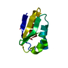
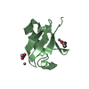
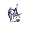
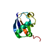
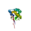
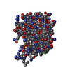
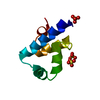
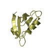
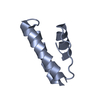
| Title | Structure of the S. venezulae BldD DNA-binding domain | ||||||
 Components Components | Putative DNA-binding protein | ||||||
 Keywords Keywords | DNA BINDING PROTEIN / BldD DNA binding domain / helix turn helix | ||||||
| Function / homology |  Function and homology information Function and homology informationDNA-binding transcription repressor activity / protein-DNA complex / sequence-specific DNA binding / transcription cis-regulatory region binding / negative regulation of DNA-templated transcription / nucleotide binding Similarity search - Function | ||||||
| Biological species | Streptomyces coelicolor A3 | ||||||
| Method |  X-RAY DIFFRACTION / SYNCHROTRON / MOLECULAR REPLACEMENT / Resolution: 2.8 Å X-RAY DIFFRACTION / SYNCHROTRON / MOLECULAR REPLACEMENT / Resolution: 2.8 Å | ||||||
 Authors Authors | schumacher, M.A. / Tschowri, N. / Buttner, M. / Brennan, R. | ||||||
 Citation Citation | Journal: Cell(Cambridge,Mass.) / Year: 2014 Title: Tetrameric c-di-GMP mediates effective transcription factor dimerization to control Streptomyces development. Authors: Tschowri, N. / Schumacher, M.A. / Schlimpert, S. / Chinnam, N.B. / Findlay, K.C. / Brennan, R.G. / Buttner, M.J. | ||||||
| History |
|
- Structure visualization
| Structure viewer | Molecule: MolmilJmol/JSmol |
|---|
- Downloads & links
-Download
| PDBx/mmCIF format | 4ob4.cif.gz | 50.3 KB | Display | PDBx/mmCIF format |
|---|---|---|---|---|
| PDB format | pdb4ob4.ent.gz | 37.2 KB | Display | PDB format |
| PDBx/mmJSON format | 4ob4.json.gz | Tree view | PDBx/mmJSON format | |
| Others |  Other downloads Other downloads |
-Validation report
| Arichive directory | https://data.pdbj.org/pub/pdb/validation_reports/ob/4ob4ftp://data.pdbj.org/pub/pdb/validation_reports/ob/4ob4 | HTTPS FTP |
|---|
-Related structure data
| Related structure data |  4oayC  4oazC  5khdC  2ewtS  4oax S: Starting model for refinement C: citing same article ( |
|---|---|
| Similar structure data |

-Links
 PDBj
PDBj


- Assembly
| Deposited unit | 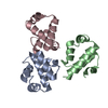
| ||||||||
|---|---|---|---|---|---|---|---|---|---|
| 1 | 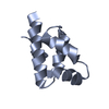
| ||||||||
| 2 | 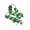
| ||||||||
| 3 | 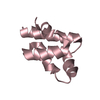
| ||||||||
| 4 | 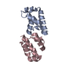
| ||||||||
| 5 | 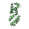
| ||||||||
| Unit cell |
|
-Components
| #1: Protein | Mass: 7883.973 Da / Num. of mol.: 3 / Fragment: DNA binding domain Source method: isolated from a genetically manipulated source Source: (gene. exp.)  Streptomyces coelicolor A3(2) (bacteria) Streptomyces coelicolor A3(2) (bacteria)Strain: ATCC 31267 / DSM 46492 / JCM 5070 / NCIMB 12804 / NRRL 8165 / MA-4680 Gene: bldD, SAV_6861, SCO1489 / Production host: #2: Water | ChemComp-HOH / |  Mass: 18.015 Da / Num. of mol.: 7 / Source method: isolated from a natural source / Formula: H2O Mass: 18.015 Da / Num. of mol.: 7 / Source method: isolated from a natural source / Formula: H2O |
|---|
-Experimental details
-Experiment
| Experiment | Method: X-RAY DIFFRACTION / Number of used crystals: 1 |
|---|
- Sample preparation
| Crystal | Density Matthews: 2.29 Å3/Da / Density % sol: 46.31 % |
|---|---|
| Crystal grow | Temperature: 298 K / Method: vapor diffusion, hanging drop / pH: 7.5 Details: 30% PEG 400, 0.1 M MgCl2, Hepes, pH 7.5, VAPOR DIFFUSION, HANGING DROP, temperature 298K |
-Data collection
| Diffraction | Mean temperature: 100 K |
|---|---|
| Diffraction source | Source: SYNCHROTRON / Site: ALS  / Beamline: 8.3.1 / Wavelength: 1 Å / Beamline: 8.3.1 / Wavelength: 1 Å |
| Detector | Type: ADSC QUANTUM 315r / Detector: CCD / Date: Mar 12, 2013 / Details: mirrors |
| Radiation | Monochromator: si(111) / Protocol: SINGLE WAVELENGTH / Monochromatic (M) / Laue (L): M / Scattering type: x-ray |
| Radiation wavelength | Wavelength: 1 Å / Relative weight: 1 |
| Reflection | Resolution: 2.8→69.256 Å / Num. obs: 5897 / % possible obs: 99.9 % / Rmerge(I) obs: 0.113 / Rsym value: 0.113 / Net I/σ(I): 12.4 |
- Processing
| Software |
| |||||||||||||||||||||||||||||||||||
|---|---|---|---|---|---|---|---|---|---|---|---|---|---|---|---|---|---|---|---|---|---|---|---|---|---|---|---|---|---|---|---|---|---|---|---|---|
| Refinement | Method to determine structure: MOLECULAR REPLACEMENT Starting model: pdb entry 2EWT Resolution: 2.8→69.256 Å / SU ML: 0.45 / σ(F): 1.38 / Phase error: 28.46 / Stereochemistry target values: ML
| |||||||||||||||||||||||||||||||||||
| Solvent computation | Shrinkage radii: 0.83 Å / VDW probe radii: 1.1 Å / Solvent model: FLAT BULK SOLVENT MODEL / Bsol: 39.906 Å2 / ksol: 0.376 e/Å3 | |||||||||||||||||||||||||||||||||||
| Displacement parameters |
| |||||||||||||||||||||||||||||||||||
| Refinement step | Cycle: LAST / Resolution: 2.8→69.256 Å
| |||||||||||||||||||||||||||||||||||
| Refine LS restraints |
| |||||||||||||||||||||||||||||||||||
| LS refinement shell |
|
