 Movie
Movie Controller


+ Open data

- Basic information
| Entry | Database: PDB / ID: 2nn4 | ||||||
|---|---|---|---|---|---|---|---|
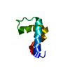
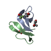
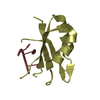
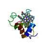
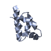
| Title | Crystal structure of Bacillus subtilis yqgQ, Pfam DUF910 | ||||||
 Components Components | Hypothetical protein yqgQ | ||||||
 Keywords Keywords | STRUCTURAL GENOMICS / UNKNOWN FUNCTION / Novel fold / pfam:DUF910 / 10278a / PSI-2 / Protein Structure Initiative / New York SGX Research Center for Structural Genomics / NYSGXRC | ||||||
| Function / homology | YqgQ-like / YqgQ-like / YqgQ-like superfamily / YqgQ-like / Helix Hairpins / Orthogonal Bundle / Mainly Alpha / Uncharacterized protein YqgQ Function and homology information Function and homology information | ||||||
| Biological species |  | ||||||
| Method |  X-RAY DIFFRACTION / SYNCHROTRON / SAD / Resolution: 2.1 Å X-RAY DIFFRACTION / SYNCHROTRON / SAD / Resolution: 2.1 Å | ||||||
 Authors Authors | Damodharan, L. / Eswaramoorthy, S. / Burley, S.K. / Swaminathan, S. / New York SGX Research Center for Structural Genomics (NYSGXRC) | ||||||
 Citation Citation | Journal: Acta Crystallogr.,Sect.F / Year: 2010 Title: Structure of YqgQ protein from Bacillus subtilis, a conserved hypothetical protein. Authors: Lakshminarasimhan, D. / Eswaramoorthy, S. / Burley, S.K. / Swaminathan, S. | ||||||
| History |
|
- Structure visualization
| Structure viewer | Molecule: MolmilJmol/JSmol |
|---|
- Downloads & links
-Download
| PDBx/mmCIF format | 2nn4.cif.gz | 50.2 KB | Display | PDBx/mmCIF format |
|---|---|---|---|---|
| PDB format | pdb2nn4.ent.gz | 37.1 KB | Display | PDB format |
| PDBx/mmJSON format | 2nn4.json.gz | Tree view | PDBx/mmJSON format | |
| Others |  Other downloads Other downloads |
-Validation report
| Arichive directory | https://data.pdbj.org/pub/pdb/validation_reports/nn/2nn4ftp://data.pdbj.org/pub/pdb/validation_reports/nn/2nn4 | HTTPS FTP |
|---|
-Related structure data
| Similar structure data | |
|---|---|
| Other databases |


-Links
 PDBj
PDBj- Assembly
| Deposited unit | 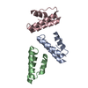
| ||||||||
|---|---|---|---|---|---|---|---|---|---|
| 1 | 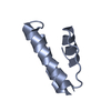
| ||||||||
| 2 | 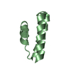
| ||||||||
| 3 | 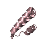
| ||||||||
| Unit cell |
|
-Components
| #1: Protein | Mass: 8701.916 Da / Num. of mol.: 3 Source method: isolated from a genetically manipulated source Source: (gene. exp.) #2: Water | ChemComp-HOH / |  Mass: 18.015 Da / Num. of mol.: 47 / Source method: isolated from a natural source / Formula: H2O Mass: 18.015 Da / Num. of mol.: 47 / Source method: isolated from a natural source / Formula: H2O |
|---|
-Experimental details
-Experiment
| Experiment | Method: X-RAY DIFFRACTION / Number of used crystals: 1 |
|---|
- Sample preparation
| Crystal | Density Matthews: 2.07 Å3/Da / Density % sol: 40.71 % |
|---|---|
| Crystal grow | Temperature: 278 K / Method: vapor diffusion, sitting drop / pH: 8.2 Details: 0.066M sodium di-hydrogen phophate, 3.385M di-potassium hydrogen phosphate, pH 8.2, VAPOR DIFFUSION, SITTING DROP, temperature 278K |
-Data collection
| Diffraction | Mean temperature: 100 K |
|---|---|
| Diffraction source | Source: SYNCHROTRON / Site: NSLS  / Beamline: X25 / Wavelength: 0.9801 / Beamline: X25 / Wavelength: 0.9801 |
| Detector | Type: ADSC QUANTUM 315 / Detector: CCD / Date: Oct 14, 2006 / Details: Mirrors |
| Radiation | Monochromator: Si(111) / Protocol: SINGLE WAVELENGTH / Monochromatic (M) / Laue (L): M / Scattering type: x-ray |
| Radiation wavelength | Wavelength: 0.9801 Å / Relative weight: 1 |
| Reflection | Resolution: 2.1→50 Å / Num. all: 12344 / Num. obs: 12344 / % possible obs: 97.5 % / Observed criterion σ(I): 0 / Redundancy: 6.5 % / Biso Wilson estimate: 17.7 Å2 / Rmerge(I) obs: 0.08 / Net I/σ(I): 12.2 |
| Reflection shell | Resolution: 2.1→2.18 Å / Redundancy: 3.9 % / Rmerge(I) obs: 0.37 / Mean I/σ(I) obs: 2.8 / Num. unique all: 1075 / % possible all: 84.7 |
- Processing
| Software |
| ||||||||||||||||||||||||||||||||||||
|---|---|---|---|---|---|---|---|---|---|---|---|---|---|---|---|---|---|---|---|---|---|---|---|---|---|---|---|---|---|---|---|---|---|---|---|---|---|
| Refinement | Method to determine structure: SAD / Resolution: 2.1→47.59 Å / Rfactor Rfree error: 0.009 / Data cutoff high absF: 127065.32 / Data cutoff low absF: 0 / Isotropic thermal model: RESTRAINED / Cross valid method: THROUGHOUT / σ(F): 0 / Stereochemistry target values: Engh & Huber Details: the missing residues listed in Remark 465 are due to lack of electron density.
| ||||||||||||||||||||||||||||||||||||
| Solvent computation | Solvent model: FLAT MODEL / Bsol: 51.1294 Å2 / ksol: 0.390188 e/Å3 | ||||||||||||||||||||||||||||||||||||
| Displacement parameters | Biso mean: 38.1 Å2
| ||||||||||||||||||||||||||||||||||||
| Refine analyze |
| ||||||||||||||||||||||||||||||||||||
| Refinement step | Cycle: LAST / Resolution: 2.1→47.59 Å
| ||||||||||||||||||||||||||||||||||||
| Refine LS restraints |
| ||||||||||||||||||||||||||||||||||||
| LS refinement shell | Resolution: 2.1→2.23 Å / Rfactor Rfree error: 0.033 / Total num. of bins used: 6
| ||||||||||||||||||||||||||||||||||||
| Xplor file |
|
