 Movie
Movie Controller


+ Open data

- Basic information
| Entry | Database: PDB / ID: 5x3p | ||||||
|---|---|---|---|---|---|---|---|
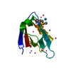
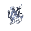
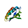
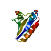
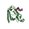
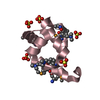
| Title | Crystal structure of the UBX domain of human UBXD7 | ||||||
 Components Components | UBX domain-containing protein 7 | ||||||
 Keywords Keywords | PROTEIN BINDING / UBXD7 / p97 | ||||||
| Function / homology |  Function and homology information Function and homology informationVCP-NPL4-UFD1 AAA ATPase complex / ubiquitin binding / KEAP1-NFE2L2 pathway / Neddylation / RNA polymerase II-specific DNA-binding transcription factor binding / proteasome-mediated ubiquitin-dependent protein catabolic process / ubiquitin protein ligase binding / nucleoplasm / nucleus / cytosol Similarity search - Function | ||||||
| Biological species |  Homo sapiens (human) Homo sapiens (human) | ||||||
| Method |  X-RAY DIFFRACTION / SYNCHROTRON / SAD / Resolution: 1.999 Å X-RAY DIFFRACTION / SYNCHROTRON / SAD / Resolution: 1.999 Å | ||||||
 Authors Authors | Jiang, T. / Li, Z. / Wang, Y. / Xu, M. | ||||||
 Citation Citation | Journal: Biochem. Biophys. Res. Commun. / Year: 2017 Title: Crystal structures of the UBX domain of human UBXD7 and its complex with p97 ATPase Authors: Li, Z.H. / Wang, Y. / Xu, M. / Jiang, T. | ||||||
| History |
|
- Structure visualization
| Structure viewer | Molecule: MolmilJmol/JSmol |
|---|
- Downloads & links
-Download
| PDBx/mmCIF format | 5x3p.cif.gz | 112 KB | Display | PDBx/mmCIF format |
|---|---|---|---|---|
| PDB format | pdb5x3p.ent.gz | 87.2 KB | Display | PDB format |
| PDBx/mmJSON format | 5x3p.json.gz | Tree view | PDBx/mmJSON format | |
| Others |  Other downloads Other downloads |
-Validation report
| Arichive directory | https://data.pdbj.org/pub/pdb/validation_reports/x3/5x3pftp://data.pdbj.org/pub/pdb/validation_reports/x3/5x3p | HTTPS FTP |
|---|
-Related structure data


-Links
 PDBj
PDBj

- Assembly
| Deposited unit | 
| ||||||||
|---|---|---|---|---|---|---|---|---|---|
| 1 | 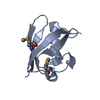
| ||||||||
| 2 | 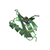
| ||||||||
| 3 | 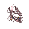
| ||||||||
| Unit cell |
|
-Components
| #1: Protein | Mass: 9633.721 Da / Num. of mol.: 3 / Fragment: UNP residues 410-489 / Mutation: L472M Source method: isolated from a genetically manipulated source Source: (gene. exp.) Homo sapiens (human) / Gene: UBXN7, KIAA0794, UBXD7 / Production host:  #2: Water | ChemComp-HOH / |  Mass: 18.015 Da / Num. of mol.: 165 / Source method: isolated from a natural source / Formula: H2O Mass: 18.015 Da / Num. of mol.: 165 / Source method: isolated from a natural source / Formula: H2OHas protein modification | Y | |
|---|
-Experimental details
-Experiment
| Experiment | Method: X-RAY DIFFRACTION / Number of used crystals: 1 |
|---|
- Sample preparation
| Crystal | Density Matthews: 1.96 Å3/Da / Density % sol: 37.22 % |
|---|---|
| Crystal grow | Temperature: 289 K / Method: vapor diffusion Details: 0.1M Tris pH 8.8, 35% v/v Polyethylene glycol 400, 0.1M LiCl |
-Data collection
| Diffraction | Mean temperature: 100 K |
|---|---|
| Diffraction source | Source: SYNCHROTRON / Site: SSRF  / Beamline: BL17U / Wavelength: 0.9792 Å / Beamline: BL17U / Wavelength: 0.9792 Å |
| Detector | Type: ADSC QUANTUM 315r / Detector: CCD / Date: Sep 9, 2016 |
| Radiation | Protocol: SINGLE WAVELENGTH / Monochromatic (M) / Laue (L): M / Scattering type: x-ray |
| Radiation wavelength | Wavelength: 0.9792 Å / Relative weight: 1 |
| Reflection | Resolution: 1.99→38.24 Å / Num. obs: 14643 / % possible obs: 99.8 % / Redundancy: 5.7 % / Rmerge(I) obs: 0.108 / Rpim(I) all: 0.048 / Net I/σ(I): 16.2 |
- Processing
| Software |
| ||||||||||||||||||||||||||||||||||||||||||
|---|---|---|---|---|---|---|---|---|---|---|---|---|---|---|---|---|---|---|---|---|---|---|---|---|---|---|---|---|---|---|---|---|---|---|---|---|---|---|---|---|---|---|---|
| Refinement | Method to determine structure: SAD / Resolution: 1.999→38.24 Å / SU ML: 0.23 / Cross valid method: FREE R-VALUE / σ(F): 1.38 / Phase error: 24.14
| ||||||||||||||||||||||||||||||||||||||||||
| Solvent computation | Shrinkage radii: 0.9 Å / VDW probe radii: 1.11 Å | ||||||||||||||||||||||||||||||||||||||||||
| Refinement step | Cycle: LAST / Resolution: 1.999→38.24 Å
| ||||||||||||||||||||||||||||||||||||||||||
| Refine LS restraints |
| ||||||||||||||||||||||||||||||||||||||||||
| LS refinement shell |
| ||||||||||||||||||||||||||||||||||||||||||
| Refinement TLS params. | Method: refined / Origin x: 16.5605 Å / Origin y: 75.6638 Å / Origin z: 7.0928 Å
| ||||||||||||||||||||||||||||||||||||||||||
| Refinement TLS group | Selection details: all |
