 Movie
Movie Controller

[English] 日本語
 Yorodumi
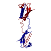
- PDB-6lay: Domain-swapped dimer structure of a Single-chain Monellin loop1-d... -
+ Open data

- Basic information
| Entry | Database: PDB / ID: 6lay | ||||||
|---|---|---|---|---|---|---|---|
| Title | Domain-swapped dimer structure of a Single-chain Monellin loop1-delta4-QVVAG mutant | ||||||
 Components Components | Monellin chain B,Monellin chain A | ||||||
 Keywords Keywords | PLANT PROTEIN / Monomer / L1 mutant | ||||||
| Function / homology | Monellin, A chain / Monellin, A chain superfamily / Monellin, B chain / : / Monellin / Monellin / Cystatin superfamily / Monellin chain A / Monellin chain B Function and homology information Function and homology information | ||||||
| Biological species |  Dioscoreophyllum cumminsii (serendipity berry) Dioscoreophyllum cumminsii (serendipity berry) | ||||||
| Method |  X-RAY DIFFRACTION / MOLECULAR REPLACEMENT / molecular replacement / Resolution: 3.001 Å X-RAY DIFFRACTION / MOLECULAR REPLACEMENT / molecular replacement / Resolution: 3.001 Å | ||||||
 Authors Authors | Manjula, R. / Subramanian, R. / Gosavi, S. | ||||||
 Citation Citation | Journal: To Be Published Title: Domain-swapped dimer structure of a Single-chain Monellin loop1-delta4-QVVAG mutant Authors: Manjula, R. / Subramanian, R. / Gosavi, S. | ||||||
| History |
|
- Structure visualization
| Structure viewer | Molecule: MolmilJmol/JSmol |
|---|
- Downloads & links
-Download
| PDBx/mmCIF format | 6lay.cif.gz | 49.4 KB | Display | PDBx/mmCIF format |
|---|---|---|---|---|
| PDB format | pdb6lay.ent.gz | 34.4 KB | Display | PDB format |
| PDBx/mmJSON format | 6lay.json.gz | Tree view | PDBx/mmJSON format | |
| Others |  Other downloads Other downloads |
-Validation report
| Arichive directory | https://data.pdbj.org/pub/pdb/validation_reports/la/6layftp://data.pdbj.org/pub/pdb/validation_reports/la/6lay | HTTPS FTP |
|---|
-Related structure data
| Related structure data |  2o9uS S: Starting model for refinement |
|---|---|
| Similar structure data |


-Links
 PDBj
PDBj
- Assembly
| Deposited unit | 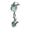
| ||||||||
|---|---|---|---|---|---|---|---|---|---|
| 1 |
| ||||||||
| Unit cell |
|
-Components
| #1: Protein | Mass: 10840.429 Da / Num. of mol.: 2 Source method: isolated from a genetically manipulated source Details: The fusion protein of Monellin chain B, LINKER and Monellin chain A Source: (gene. exp.) Dioscoreophyllum cumminsii (serendipity berry)Production host:  |
|---|
-Experimental details
-Experiment
| Experiment | Method: X-RAY DIFFRACTION / Number of used crystals: 1 |
|---|
- Sample preparation
| Crystal | Density Matthews: 2.33 Å3/Da / Density % sol: 47.1 % |
|---|---|
| Crystal grow | Temperature: 293 K / Method: vapor diffusion, hanging drop / pH: 8.5 Details: 20% PEG 3350, 0.2mM sodium acetate ,Tris HCl pH 8.5 |
-Data collection
| Diffraction | Mean temperature: 80 K / Serial crystal experiment: N | ||||||||||||||||||||||||||||||
|---|---|---|---|---|---|---|---|---|---|---|---|---|---|---|---|---|---|---|---|---|---|---|---|---|---|---|---|---|---|---|---|
| Diffraction source | Source: ROTATING ANODE / Type: RIGAKU FR-X / Wavelength: 1.5478 Å | ||||||||||||||||||||||||||||||
| Detector | Type: RIGAKU RAXIS IV++ / Detector: IMAGE PLATE / Date: Apr 8, 2019 | ||||||||||||||||||||||||||||||
| Radiation | Protocol: SINGLE WAVELENGTH / Monochromatic (M) / Laue (L): M / Scattering type: x-ray | ||||||||||||||||||||||||||||||
| Radiation wavelength | Wavelength: 1.5478 Å / Relative weight: 1 | ||||||||||||||||||||||||||||||
| Reflection | Resolution: 3→45.29 Å / Num. obs: 4033 / % possible obs: 100 % / Redundancy: 5.3 % / CC1/2: 0.996 / Rmerge(I) obs: 0.098 / Rpim(I) all: 0.047 / Rrim(I) all: 0.109 / Net I/σ(I): 12.5 / Num. measured all: 21231 / Scaling rejects: 6 | ||||||||||||||||||||||||||||||
| Reflection shell | Diffraction-ID: 1
|
-Phasing
| Phasing | Method: molecular replacement |
|---|
- Processing
| Software |
| ||||||||||||||||||||
|---|---|---|---|---|---|---|---|---|---|---|---|---|---|---|---|---|---|---|---|---|---|
| Refinement | Method to determine structure: MOLECULAR REPLACEMENT Starting model: 2O9U Resolution: 3.001→45.289 Å / SU ML: 0.39 / Cross valid method: THROUGHOUT / σ(F): 1.35 / Phase error: 19.73 / Stereochemistry target values: ML
| ||||||||||||||||||||
| Solvent computation | Shrinkage radii: 0.9 Å / VDW probe radii: 1.11 Å / Solvent model: FLAT BULK SOLVENT MODEL | ||||||||||||||||||||
| Displacement parameters | Biso max: 46.47 Å2 / Biso mean: 32.272 Å2 / Biso min: 21.7 Å2 | ||||||||||||||||||||
| Refinement step | Cycle: final / Resolution: 3.001→45.289 Å
| ||||||||||||||||||||
| LS refinement shell | Resolution: 3.001→3.18 Å / Rfactor Rfree error: 0
|
