 Movie
Movie Controller

[English] 日本語
 Yorodumi
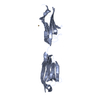
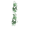
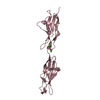
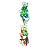
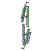
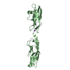
- PDB-6fwx: Chimeric titin Z1Z2-Z1Z2 tandem (Z1212) functionalized with a GRG... -
+ Open data

- Basic information
| Entry | Database: PDB / ID: 6fwx | ||||||
|---|---|---|---|---|---|---|---|
| Title | Chimeric titin Z1Z2-Z1Z2 tandem (Z1212) functionalized with a GRGDS exogenous peptide from fibronectin | ||||||
 Components Components | Titin,Titin,Titin | ||||||
 Keywords Keywords | STRUCTURAL PROTEIN / titin / Z1Z2 | ||||||
| Function / homology |  Function and homology information Function and homology informationsarcomerogenesis / titin-telethonin complex / structural molecule activity conferring elasticity / skeletal muscle myosin thick filament assembly / telethonin binding / detection of muscle stretch / : / muscle alpha-actinin binding / cardiac myofibril assembly / cardiac muscle hypertrophy ...sarcomerogenesis / titin-telethonin complex / structural molecule activity conferring elasticity / skeletal muscle myosin thick filament assembly / telethonin binding / detection of muscle stretch / : / muscle alpha-actinin binding / cardiac myofibril assembly / cardiac muscle hypertrophy / cardiac muscle tissue morphogenesis / protein kinase regulator activity / mitotic chromosome condensation / Striated Muscle Contraction / muscle filament sliding / actinin binding / M band / cardiac muscle cell development / sarcomere organization / I band / structural constituent of muscle / striated muscle thin filament / skeletal muscle thin filament assembly / skeletal muscle contraction / cardiac muscle contraction / muscle contraction / striated muscle contraction / condensed nuclear chromosome / positive regulation of protein secretion / response to calcium ion / Z disc / actin filament binding / Platelet degranulation / actin cytoskeleton / protease binding / protein tyrosine kinase activity / calmodulin binding / non-specific serine/threonine protein kinase / protein serine kinase activity / protein serine/threonine kinase activity / calcium ion binding / positive regulation of gene expression / protein kinase binding / enzyme binding / protein homodimerization activity / extracellular exosome / extracellular region / ATP binding / identical protein binding / cytosol Similarity search - Function | ||||||
| Biological species |  Homo sapiens (human) Homo sapiens (human) | ||||||
| Method |  X-RAY DIFFRACTION / SYNCHROTRON / MOLECULAR REPLACEMENT / Resolution: 3 Å X-RAY DIFFRACTION / SYNCHROTRON / MOLECULAR REPLACEMENT / Resolution: 3 Å | ||||||
 Authors Authors | Mayans, O. / Fleming, J. / Hill, C. | ||||||
| Funding support |  United Kingdom, 1items United Kingdom, 1items
| ||||||
 Citation Citation | Journal: Adv. Mater. Weinheim / Year: 2019 Title: Self-Assembling Proteins as High-Performance Substrates for Embryonic Stem Cell Self-Renewal. Authors: Hill, C.J. / Fleming, J.R. / Mousavinejad, M. / Nicholson, R. / Tzokov, S.B. / Bullough, P.A. / Bogomolovas, J. / Morgan, M.R. / Mayans, O. / Murray, P. #1: Journal: Int J Mol Sci / Year: 2019Title: The ZT Biopolymer: A Self-Assembling Protein Scaffold for Stem Cell Applications. Authors: Nesterenko, Y. / Hill, C.J. / Fleming, J.R. / Murray, P. / Mayans, O. | ||||||
| History |
|
- Structure visualization
| Structure viewer | Molecule: MolmilJmol/JSmol |
|---|
- Downloads & links
-Download
| PDBx/mmCIF format | 6fwx.cif.gz | 92.3 KB | Display | PDBx/mmCIF format |
|---|---|---|---|---|
| PDB format | pdb6fwx.ent.gz | 67 KB | Display | PDB format |
| PDBx/mmJSON format | 6fwx.json.gz | Tree view | PDBx/mmJSON format | |
| Others |  Other downloads Other downloads |
-Validation report
| Arichive directory | https://data.pdbj.org/pub/pdb/validation_reports/fw/6fwxftp://data.pdbj.org/pub/pdb/validation_reports/fw/6fwx | HTTPS FTP |
|---|
-Related structure data
| Related structure data |  2a38S S: Starting model for refinement |
|---|---|
| Similar structure data |


-Links
 PDBj
PDBj


- Assembly
| Deposited unit | 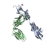
| ||||||||
|---|---|---|---|---|---|---|---|---|---|
| 1 | 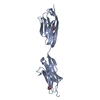
| ||||||||
| 2 | 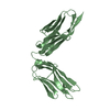
| ||||||||
| Unit cell |
|
-Components
| #1: Protein | Mass: 41934.480 Da / Num. of mol.: 2 Source method: isolated from a genetically manipulated source Source: (gene. exp.) Homo sapiens (human) / Gene: TTN / Production host:  References: UniProt: Q8WZ42, non-specific serine/threonine protein kinase #2: Chemical |   Mass: 62.068 Da / Num. of mol.: 2 / Source method: obtained synthetically / Formula: C2H6O2 Mass: 62.068 Da / Num. of mol.: 2 / Source method: obtained synthetically / Formula: C2H6O2 |
|---|
-Experimental details
-Experiment
| Experiment | Method: X-RAY DIFFRACTION / Number of used crystals: 1 |
|---|
- Sample preparation
| Crystal | Density Matthews: 2.78 Å3/Da / Density % sol: 55.71 % |
|---|---|
| Crystal grow | Temperature: 293 K / Method: vapor diffusion, sitting drop Details: 10% [v/v] isopropanol, 100 mM Na2HPO4 pH 4.2, 200 mM LiSO4 |
-Data collection
| Diffraction | Mean temperature: 100 K |
|---|---|
| Diffraction source | Source: SYNCHROTRON / Site: Diamond / Beamline: I03 / Wavelength: 0.974 Å |
| Detector | Type: DECTRIS PILATUS3 6M / Detector: PIXEL / Date: Aug 23, 2015 |
| Radiation | Protocol: SINGLE WAVELENGTH / Monochromatic (M) / Laue (L): M / Scattering type: x-ray |
| Radiation wavelength | Wavelength: 0.974 Å / Relative weight: 1 |
| Reflection | Resolution: 3→41.8 Å / Num. obs: 17993 / % possible obs: 99 % / Redundancy: 4.1 % / Net I/σ(I): 9.26 |
| Reflection shell | Resolution: 3→3.108 Å / Rmerge(I) obs: 1.803 / Mean I/σ(I) obs: 0.6 / Num. unique obs: 1813 / CC1/2: 0.162 / Rrim(I) all: 2.079 |
- Processing
| Software |
| |||||||||||||||||||||||||||||||||||||||||||||||||
|---|---|---|---|---|---|---|---|---|---|---|---|---|---|---|---|---|---|---|---|---|---|---|---|---|---|---|---|---|---|---|---|---|---|---|---|---|---|---|---|---|---|---|---|---|---|---|---|---|---|---|
| Refinement | Method to determine structure: MOLECULAR REPLACEMENT Starting model: 2A38 Resolution: 3→41.798 Å / SU ML: 0.69 / Cross valid method: FREE R-VALUE / σ(F): 1.92 / Phase error: 30.67
| |||||||||||||||||||||||||||||||||||||||||||||||||
| Solvent computation | Shrinkage radii: 0.9 Å / VDW probe radii: 1.11 Å | |||||||||||||||||||||||||||||||||||||||||||||||||
| Refinement step | Cycle: LAST / Resolution: 3→41.798 Å
| |||||||||||||||||||||||||||||||||||||||||||||||||
| Refine LS restraints |
| |||||||||||||||||||||||||||||||||||||||||||||||||
| LS refinement shell |
|
