 Movie
Movie Controller


+ Open data

- Basic information
| Entry | Database: PDB / ID: 1lxe | ||||||
|---|---|---|---|---|---|---|---|
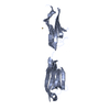
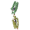
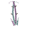
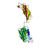
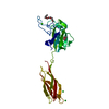
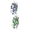
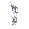
| Title | CRYSTAL STRUCTURE OF THE CATHELICIDIN MOTIF OF PROTEGRINS | ||||||
 Components Components | protegrin-3 precursor | ||||||
 Keywords Keywords | ANTIMICROBIAL PROTEIN / PROTEGRIN / CATHELICIDIN MOTIF / DISULFIDE / domain swapping | ||||||
| Function / homology |  Function and homology information Function and homology informationlipopolysaccharide binding / antimicrobial humoral immune response mediated by antimicrobial peptide / defense response to Gram-negative bacterium / defense response to Gram-positive bacterium / innate immune response / : Similarity search - Function | ||||||
| Biological species |  | ||||||
| Method |  X-RAY DIFFRACTION / FOURIER SYNTHESIS / Resolution: 2.5 Å X-RAY DIFFRACTION / FOURIER SYNTHESIS / Resolution: 2.5 Å | ||||||
 Authors Authors | Sanchez, J.F. / Hoh, F. / Strub, M.P. / Aumelas, A. / Dumas, C. | ||||||
 Citation Citation | Journal: Structure / Year: 2002 Title: Structure of the cathelicidin motif of protegrin-3 precursor: structural insights into the activation mechanism of an antimicrobial protein. Authors: Sanchez, J.F. / Hoh, F. / Strub, M.P. / Aumelas, A. / Dumas, C. | ||||||
| History |
|
- Structure visualization
| Structure viewer | Molecule: MolmilJmol/JSmol |
|---|
- Downloads & links
-Download
| PDBx/mmCIF format | 1lxe.cif.gz | 31.1 KB | Display | PDBx/mmCIF format |
|---|---|---|---|---|
| PDB format | pdb1lxe.ent.gz | 20.7 KB | Display | PDB format |
| PDBx/mmJSON format | 1lxe.json.gz | Tree view | PDBx/mmJSON format | |
| Others |  Other downloads Other downloads |
-Validation report
| Arichive directory | https://data.pdbj.org/pub/pdb/validation_reports/lx/1lxeftp://data.pdbj.org/pub/pdb/validation_reports/lx/1lxe | HTTPS FTP |
|---|
-Related structure data
| Related structure data | 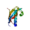 1kwiSC S: Starting model for refinement C: citing same article ( |
|---|---|
| Similar structure data |

-Links
 PDBj
PDBj
- Assembly
| Deposited unit | 
| ||||||||
|---|---|---|---|---|---|---|---|---|---|
| 1 | 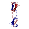
| ||||||||
| Unit cell |
| ||||||||
| Details | The second part of the biological assembly is generated by the two fold axis: Rotation : 0.5000 0.8660 0.0 0.8660 -0.5000 0.0 0.0 0.0 -1.0 Translation : -26.086 45.183 -22.580 |
-Components
| #1: Protein | Mass: 11322.741 Da / Num. of mol.: 1 / Fragment: residues 30-130 Source method: isolated from a genetically manipulated source Source: (gene. exp.)  |
|---|---|
| #2: Water | ChemComp-HOH /  Mass: 18.015 Da / Num. of mol.: 45 / Source method: isolated from a natural source / Formula: H2O Mass: 18.015 Da / Num. of mol.: 45 / Source method: isolated from a natural source / Formula: H2O |
| Has protein modification | Y |
-Experimental details
-Experiment
| Experiment | Method: X-RAY DIFFRACTION / Number of used crystals: 1 |
|---|
- Sample preparation
| Crystal | Density Matthews: 2.35 Å3/Da / Density % sol: 47.62 % | ||||||||||||||||||||||||
|---|---|---|---|---|---|---|---|---|---|---|---|---|---|---|---|---|---|---|---|---|---|---|---|---|---|
| Crystal grow | Temperature: 291 K / Method: vapor diffusion, hanging drop / pH: 3.5 Details: AMMONIUM SULFATE, SODIUM ACETATE, pH 3.5, VAPOR DIFFUSION, HANGING DROP, temperature 291K | ||||||||||||||||||||||||
| Crystal grow | *PLUS Temperature: 18 ℃ / PH range low: 3.7 / PH range high: 3.5 | ||||||||||||||||||||||||
| Components of the solutions | *PLUS
|
-Data collection
| Diffraction | Mean temperature: 291 K |
|---|---|
| Diffraction source | Source: ROTATING ANODE / Type: RIGAKU RU200 / Wavelength: 1.5418 Å |
| Detector | Type: MARRESEARCH / Detector: IMAGE PLATE / Date: Jan 15, 2002 |
| Radiation | Monochromator: graphite / Protocol: SINGLE WAVELENGTH / Monochromatic (M) / Laue (L): M / Scattering type: x-ray |
| Radiation wavelength | Wavelength: 1.5418 Å / Relative weight: 1 |
| Reflection | Resolution: 2.5→27.1 Å / Num. all: 3922 / Num. obs: 3922 / % possible obs: 91.3 % / Observed criterion σ(F): 0 / Observed criterion σ(I): 0 / Biso Wilson estimate: 43.8 Å2 / Rmerge(I) obs: 0.092 / Rsym value: 0.092 / Net I/σ(I): 18.5 |
| Reflection shell | Resolution: 2.5→2.66 Å / Rmerge(I) obs: 0.241 / Mean I/σ(I) obs: 4.6 / Num. unique all: 541 / Rsym value: 0.241 / % possible all: 91.3 |
| Reflection | *PLUS % possible obs: 93.3 % / Num. measured all: 37505 |
| Reflection shell | *PLUS % possible obs: 92 % |
- Processing
| Software |
| ||||||||||||||||||||||||||||||||||||||||||||||||||||||||||||||||||||||||||||||||
|---|---|---|---|---|---|---|---|---|---|---|---|---|---|---|---|---|---|---|---|---|---|---|---|---|---|---|---|---|---|---|---|---|---|---|---|---|---|---|---|---|---|---|---|---|---|---|---|---|---|---|---|---|---|---|---|---|---|---|---|---|---|---|---|---|---|---|---|---|---|---|---|---|---|---|---|---|---|---|---|---|---|
| Refinement | Method to determine structure: FOURIER SYNTHESIS Starting model: PDB ENTRY 1KWI Resolution: 2.5→27.1 Å / Rfactor Rfree error: 0.014 / Data cutoff high absF: 1799954.7 / Data cutoff high rms absF: 1799954.7 / Isotropic thermal model: RESTRAINED / Cross valid method: THROUGHOUT / σ(F): 0 / σ(I): 0 / Stereochemistry target values: Engh & Huber
| ||||||||||||||||||||||||||||||||||||||||||||||||||||||||||||||||||||||||||||||||
| Solvent computation | Solvent model: FLAT MODEL / Bsol: 104.394 Å2 / ksol: 0.429523 e/Å3 | ||||||||||||||||||||||||||||||||||||||||||||||||||||||||||||||||||||||||||||||||
| Displacement parameters | Biso mean: 33.6 Å2
| ||||||||||||||||||||||||||||||||||||||||||||||||||||||||||||||||||||||||||||||||
| Refine analyze |
| ||||||||||||||||||||||||||||||||||||||||||||||||||||||||||||||||||||||||||||||||
| Refinement step | Cycle: LAST / Resolution: 2.5→27.1 Å
| ||||||||||||||||||||||||||||||||||||||||||||||||||||||||||||||||||||||||||||||||
| Refine LS restraints |
| ||||||||||||||||||||||||||||||||||||||||||||||||||||||||||||||||||||||||||||||||
| LS refinement shell | Resolution: 2.5→2.66 Å / Rfactor Rfree error: 0.033 / Total num. of bins used: 6
| ||||||||||||||||||||||||||||||||||||||||||||||||||||||||||||||||||||||||||||||||
| Xplor file |
| ||||||||||||||||||||||||||||||||||||||||||||||||||||||||||||||||||||||||||||||||
| Refinement | *PLUS Lowest resolution: 27.1 Å | ||||||||||||||||||||||||||||||||||||||||||||||||||||||||||||||||||||||||||||||||
| Solvent computation | *PLUS | ||||||||||||||||||||||||||||||||||||||||||||||||||||||||||||||||||||||||||||||||
| Displacement parameters | *PLUS | ||||||||||||||||||||||||||||||||||||||||||||||||||||||||||||||||||||||||||||||||
| Refine LS restraints | *PLUS
|
