 Movie
Movie Controller

[English] 日本語
 Yorodumi
- PDB-6l3p: Crystal strcuture of Feruloyl-CoA hydratase lyase(FCHL) complexed... -
+ Open data

- Basic information
| Entry | Database: PDB / ID: 6l3p | ||||||
|---|---|---|---|---|---|---|---|
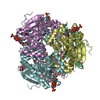
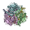
| Title | Crystal strcuture of Feruloyl-CoA hydratase lyase(FCHL) complexed with CoA | ||||||
 Components Components | Hydroxycinnamoyl-CoA hydratase-lyase | ||||||
 Keywords Keywords | LYASE / Feruloyl-CoA hydratae lyase | ||||||
| Function / homology |  Function and homology information Function and homology informationvanillin synthase / trans-feruloyl-CoA hydratase / isoprenoid catabolic process / lyase activity Similarity search - Function | ||||||
| Biological species |  Pseudomonas putida KT2440 (bacteria) Pseudomonas putida KT2440 (bacteria) | ||||||
| Method |  X-RAY DIFFRACTION / SYNCHROTRON / MOLECULAR REPLACEMENT / molecular replacement / Resolution: 2.5 Å X-RAY DIFFRACTION / SYNCHROTRON / MOLECULAR REPLACEMENT / molecular replacement / Resolution: 2.5 Å | ||||||
 Authors Authors | Seok, J. / Seo, H. / Kim, K.-J. | ||||||
 Citation Citation | Journal: to be published Title: Kinetic and structural analysis for bioproduction of vanillin by feruloyl-CoA hydratase/lyase from Pseudomonas putida KT2440 Authors: Seok, J. / Seo, H. / Kim, K.-J. | ||||||
| History |
|
- Structure visualization
| Structure viewer | Molecule: MolmilJmol/JSmol |
|---|
- Downloads & links
-Download
| PDBx/mmCIF format | 6l3p.cif.gz | 113.6 KB | Display | PDBx/mmCIF format |
|---|---|---|---|---|
| PDB format | pdb6l3p.ent.gz | 87 KB | Display | PDB format |
| PDBx/mmJSON format | 6l3p.json.gz | Tree view | PDBx/mmJSON format | |
| Others |  Other downloads Other downloads |
-Validation report
| Arichive directory | https://data.pdbj.org/pub/pdb/validation_reports/l3/6l3pftp://data.pdbj.org/pub/pdb/validation_reports/l3/6l3p | HTTPS FTP |
|---|
-Related structure data
| Related structure data | 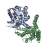 6l3oC  2j5iS S: Starting model for refinement C: citing same article ( |
|---|---|
| Similar structure data |


-Links
 PDBj
PDBj


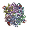
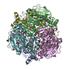
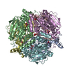
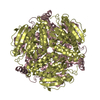
- Assembly
| Deposited unit | 
| ||||||||
|---|---|---|---|---|---|---|---|---|---|
| 1 | 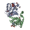
| ||||||||
| Unit cell |
|
-Components
| #1: Protein | Mass: 32123.629 Da / Num. of mol.: 2 Source method: isolated from a genetically manipulated source Source: (gene. exp.) Pseudomonas putida KT2440 (bacteria) / Strain: ATCC 47054 / DSM 6125 / NCIMB 11950 / KT2440 / Gene: PP_3358 / Plasmid: pET30a / Production host: References: UniProt: Q88HJ8, vanillin synthase, trans-feruloyl-CoA hydratase #2: Chemical |   Mass: 767.534 Da / Num. of mol.: 2 / Source method: obtained synthetically / Formula: C21H36N7O16P3S / Feature type: SUBJECT OF INVESTIGATION Mass: 767.534 Da / Num. of mol.: 2 / Source method: obtained synthetically / Formula: C21H36N7O16P3S / Feature type: SUBJECT OF INVESTIGATION#3: Chemical | ChemComp-GOL /   Mass: 92.094 Da / Num. of mol.: 5 / Source method: obtained synthetically / Formula: C3H8O3 Mass: 92.094 Da / Num. of mol.: 5 / Source method: obtained synthetically / Formula: C3H8O3#4: Water | ChemComp-HOH / |  Mass: 18.015 Da / Num. of mol.: 26 / Source method: isolated from a natural source / Formula: H2O Mass: 18.015 Da / Num. of mol.: 26 / Source method: isolated from a natural source / Formula: H2OHas ligand of interest | Y | |
|---|
-Experimental details
-Experiment
| Experiment | Method: X-RAY DIFFRACTION / Number of used crystals: 1 |
|---|
- Sample preparation
| Crystal | Density Matthews: 3.4 Å3/Da / Density % sol: 63.83 % |
|---|---|
| Crystal grow | Temperature: 293 K / Method: vapor diffusion, hanging drop / pH: 4.2 / Details: 25% propanediol, PEG3000, Glycerol |
-Data collection
| Diffraction | Mean temperature: 100 K / Serial crystal experiment: N |
|---|---|
| Diffraction source | Source: SYNCHROTRON / Site: PAL/PLS  / Beamline: 7A (6B, 6C1) / Wavelength: 0.97934 Å / Beamline: 7A (6B, 6C1) / Wavelength: 0.97934 Å |
| Detector | Type: ADSC QUANTUM 270 / Detector: CCD / Date: May 17, 2019 |
| Radiation | Monochromator: Double Crystal Monochromator / Protocol: SINGLE WAVELENGTH / Monochromatic (M) / Laue (L): M / Scattering type: x-ray |
| Radiation wavelength | Wavelength: 0.97934 Å / Relative weight: 1 |
| Reflection | Resolution: 2.5→50 Å / Num. obs: 29931 / % possible obs: 98.5 % / Redundancy: 11.2 % / CC1/2: 0.995 / Rpim(I) all: 0.022 / Rrim(I) all: 0.084 / Net I/σ(I): 38.3 |
| Reflection shell | Resolution: 2.5→2.56 Å / Num. unique obs: 2038 / CC1/2: 0.935 |
-Phasing
| Phasing | Method: molecular replacement |
|---|
- Processing
| Software |
| ||||||||||||||||||||||||||||||||||||||||||||||||||||||||||||
|---|---|---|---|---|---|---|---|---|---|---|---|---|---|---|---|---|---|---|---|---|---|---|---|---|---|---|---|---|---|---|---|---|---|---|---|---|---|---|---|---|---|---|---|---|---|---|---|---|---|---|---|---|---|---|---|---|---|---|---|---|---|
| Refinement | Method to determine structure: MOLECULAR REPLACEMENT Starting model: 2j5i Resolution: 2.5→31.52 Å / Cor.coef. Fo:Fc: 0.926 / Cor.coef. Fo:Fc free: 0.884 / SU B: 11.473 / SU ML: 0.243 / Cross valid method: THROUGHOUT / σ(F): 0 / ESU R: 0.342 / ESU R Free: 0.273 Details: HYDROGENS HAVE BEEN ADDED IN THE RIDING POSITIONS U VALUES : REFINED INDIVIDUALLY
| ||||||||||||||||||||||||||||||||||||||||||||||||||||||||||||
| Solvent computation | Ion probe radii: 0.8 Å / Shrinkage radii: 0.8 Å / VDW probe radii: 1.2 Å | ||||||||||||||||||||||||||||||||||||||||||||||||||||||||||||
| Displacement parameters | Biso max: 129.06 Å2 / Biso mean: 47.723 Å2 / Biso min: 11.58 Å2
| ||||||||||||||||||||||||||||||||||||||||||||||||||||||||||||
| Refinement step | Cycle: final / Resolution: 2.5→31.52 Å
| ||||||||||||||||||||||||||||||||||||||||||||||||||||||||||||
| Refine LS restraints |
| ||||||||||||||||||||||||||||||||||||||||||||||||||||||||||||
| LS refinement shell | Resolution: 2.502→2.567 Å / Rfactor Rfree error: 0 / Total num. of bins used: 20
|
