 Movie
Movie Controller


+ Open data

- Basic information
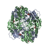
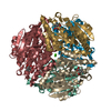
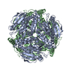
| Entry | Database: PDB / ID: 2j5i | ||||||
|---|---|---|---|---|---|---|---|
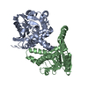
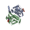
| Title | Crystal Structure of Hydroxycinnamoyl-CoA Hydratase-Lyase | ||||||
 Components Components | (P-HYDROXYCINNAMOYL COA HYDRATASE/LYASE) x 3 | ||||||
 Keywords Keywords | LYASE / VANILLIN / ALDOLASE / CROTONASE / HYDRATASE / COENZYME-A | ||||||
| Function / homology |  Function and homology information Function and homology informationferuloyl-CoA hydratase/lyase / feruloyl-CoA hydratase/lyase activity / isoprenoid catabolic process Similarity search - Function | ||||||
| Biological species |  PSEUDOMONAS FLUORESCENS (bacteria) PSEUDOMONAS FLUORESCENS (bacteria) | ||||||
| Method |  X-RAY DIFFRACTION / SYNCHROTRON / MOLECULAR REPLACEMENT / Resolution: 1.8 Å X-RAY DIFFRACTION / SYNCHROTRON / MOLECULAR REPLACEMENT / Resolution: 1.8 Å | ||||||
 Authors Authors | Leonard, P.M. / Brzozowski, A.M. / Lebedev, A. / Marshall, C.M. / Smith, D.J. / Verma, C.S. / Walton, N.J. / Grogan, G. | ||||||
 Citation Citation | Journal: Acta Crystallogr.,Sect.D / Year: 2006 Title: The 1.8 A Resolution Structure of Hydroxycinnamoyl- Coenzyme a Hydratase-Lyase (Hchl) from Pseudomonas Fluorescens, an Enzyme that Catalyses the Transformation of Feruloyl-Coenzyme a to Vanillin. Authors: Leonard, P.M. / Brzozowski, A.M. / Lebedev, A. / Marshall, C.M. / Smith, D.J. / Verma, C.S. / Walton, N.J. / Grogan, G. | ||||||
| History |
|
- Structure visualization
| Structure viewer | Molecule: MolmilJmol/JSmol |
|---|
- Downloads & links
-Download
| PDBx/mmCIF format | 2j5i.cif.gz | 611.9 KB | Display | PDBx/mmCIF format |
|---|---|---|---|---|
| PDB format | pdb2j5i.ent.gz | 503.5 KB | Display | PDB format |
| PDBx/mmJSON format | 2j5i.json.gz | Tree view | PDBx/mmJSON format | |
| Others |  Other downloads Other downloads |
-Validation report
| Arichive directory | https://data.pdbj.org/pub/pdb/validation_reports/j5/2j5iftp://data.pdbj.org/pub/pdb/validation_reports/j5/2j5i | HTTPS FTP |
|---|
-Related structure data
| Related structure data |  1uiyS S: Starting model for refinement |
|---|---|
| Similar structure data |

-Links
 PDBj
PDBj- Assembly
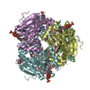
| Deposited unit | 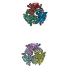
| |||||||||||||||||||||||||||||||||||||||||||||||||||||||||||||||||||||||||||||||||||||||||||||||||||||||||||||||||||||
|---|---|---|---|---|---|---|---|---|---|---|---|---|---|---|---|---|---|---|---|---|---|---|---|---|---|---|---|---|---|---|---|---|---|---|---|---|---|---|---|---|---|---|---|---|---|---|---|---|---|---|---|---|---|---|---|---|---|---|---|---|---|---|---|---|---|---|---|---|---|---|---|---|---|---|---|---|---|---|---|---|---|---|---|---|---|---|---|---|---|---|---|---|---|---|---|---|---|---|---|---|---|---|---|---|---|---|---|---|---|---|---|---|---|---|---|---|---|---|
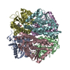
| 1 | 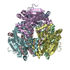
| |||||||||||||||||||||||||||||||||||||||||||||||||||||||||||||||||||||||||||||||||||||||||||||||||||||||||||||||||||||
| 2 | 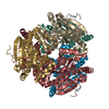
| |||||||||||||||||||||||||||||||||||||||||||||||||||||||||||||||||||||||||||||||||||||||||||||||||||||||||||||||||||||
| Unit cell |
| |||||||||||||||||||||||||||||||||||||||||||||||||||||||||||||||||||||||||||||||||||||||||||||||||||||||||||||||||||||
| Noncrystallographic symmetry (NCS) | NCS domain:
NCS domain segments:
|
-Components
| #1: Protein | Mass: 31012.637 Da / Num. of mol.: 1 Source method: isolated from a genetically manipulated source Source: (gene. exp.) PSEUDOMONAS FLUORESCENS (bacteria) / Strain: AN103 / Description: INSITUTES OF FOOD RESEARCH, NORWICH U.K. / Plasmid: PFI3009 / Production host: | ||||
|---|---|---|---|---|---|
| #2: Protein | Mass: 31044.613 Da / Num. of mol.: 10 Source method: isolated from a genetically manipulated source Source: (gene. exp.) PSEUDOMONAS FLUORESCENS (bacteria) / Strain: AN103 / Description: INSITUTES OF FOOD RESEARCH, NORWICH U.K. / Plasmid: PFI3009 / Production host: #3: Protein | | Mass: 31043.629 Da / Num. of mol.: 1 Source method: isolated from a genetically manipulated source Source: (gene. exp.) PSEUDOMONAS FLUORESCENS (bacteria) / Strain: AN103 / Description: INSITUTES OF FOOD RESEARCH, NORWICH U.K. / Plasmid: PFI3009 / Production host: #4: Water | ChemComp-HOH / |  Mass: 18.015 Da / Num. of mol.: 1861 / Source method: isolated from a natural source / Formula: H2O Mass: 18.015 Da / Num. of mol.: 1861 / Source method: isolated from a natural source / Formula: H2O |
-Experimental details
-Experiment
| Experiment | Method: X-RAY DIFFRACTION / Number of used crystals: 1 |
|---|
- Sample preparation
| Crystal | Density Matthews: 2.42 Å3/Da / Density % sol: 49.5 % |
|---|---|
| Crystal grow | pH: 5.6 Details: PEG 20000 11% W/V PEG 550 MME 8% V/V 0.8M NA FORMATE BUTANE-1,4-DIOL 0,2% V/V 0.05M MES PH 5.6 |
-Data collection
| Diffraction | Mean temperature: 287 K |
|---|---|
| Diffraction source | Source: SYNCHROTRON / Site: ESRF  / Beamline: ID14-1 / Wavelength: 0.934 / Beamline: ID14-1 / Wavelength: 0.934 |
| Detector | Type: ADSC CCD / Detector: CCD / Date: Jan 30, 2004 |
| Radiation | Protocol: SINGLE WAVELENGTH / Monochromatic (M) / Laue (L): M / Scattering type: x-ray |
| Radiation wavelength | Wavelength: 0.934 Å / Relative weight: 1 |
| Reflection | Resolution: 1.8→30 Å / Num. obs: 308927 / % possible obs: 100 % / Observed criterion σ(I): 0 / Redundancy: 4.2 % / Rmerge(I) obs: 0.04 / Net I/σ(I): 21.9 |
| Reflection shell | Resolution: 1.8→1.85 Å / Redundancy: 4.2 % / Rmerge(I) obs: 0.33 / Mean I/σ(I) obs: 3.3 / % possible all: 100 |
- Processing
| Software |
| ||||||||||||||||||||||||||||||||||||||||||||||||||||||||||||||||||||||||||||||||||||||||||||||||||||||||||||||||||||||||||||||||||||||||||||||||||||||||||||||||||||||||||||||||||||||
|---|---|---|---|---|---|---|---|---|---|---|---|---|---|---|---|---|---|---|---|---|---|---|---|---|---|---|---|---|---|---|---|---|---|---|---|---|---|---|---|---|---|---|---|---|---|---|---|---|---|---|---|---|---|---|---|---|---|---|---|---|---|---|---|---|---|---|---|---|---|---|---|---|---|---|---|---|---|---|---|---|---|---|---|---|---|---|---|---|---|---|---|---|---|---|---|---|---|---|---|---|---|---|---|---|---|---|---|---|---|---|---|---|---|---|---|---|---|---|---|---|---|---|---|---|---|---|---|---|---|---|---|---|---|---|---|---|---|---|---|---|---|---|---|---|---|---|---|---|---|---|---|---|---|---|---|---|---|---|---|---|---|---|---|---|---|---|---|---|---|---|---|---|---|---|---|---|---|---|---|---|---|---|---|
| Refinement | Method to determine structure: MOLECULAR REPLACEMENT Starting model: PDB ENTRY 1UIY Resolution: 1.8→111.8 Å / Cor.coef. Fo:Fc: 0.965 / Cor.coef. Fo:Fc free: 0.949 / Cross valid method: THROUGHOUT / ESU R: 0.117 / ESU R Free: 0.115 / Stereochemistry target values: MAXIMUM LIKELIHOOD / Details: HYDROGENS HAVE BEEN ADDED IN THE RIDING POSITIONS.
| ||||||||||||||||||||||||||||||||||||||||||||||||||||||||||||||||||||||||||||||||||||||||||||||||||||||||||||||||||||||||||||||||||||||||||||||||||||||||||||||||||||||||||||||||||||||
| Solvent computation | Ion probe radii: 0.8 Å / Shrinkage radii: 0.8 Å / VDW probe radii: 1.2 Å / Solvent model: MASK | ||||||||||||||||||||||||||||||||||||||||||||||||||||||||||||||||||||||||||||||||||||||||||||||||||||||||||||||||||||||||||||||||||||||||||||||||||||||||||||||||||||||||||||||||||||||
| Displacement parameters | Biso mean: 21.75 Å2
| ||||||||||||||||||||||||||||||||||||||||||||||||||||||||||||||||||||||||||||||||||||||||||||||||||||||||||||||||||||||||||||||||||||||||||||||||||||||||||||||||||||||||||||||||||||||
| Refinement step | Cycle: LAST / Resolution: 1.8→111.8 Å
| ||||||||||||||||||||||||||||||||||||||||||||||||||||||||||||||||||||||||||||||||||||||||||||||||||||||||||||||||||||||||||||||||||||||||||||||||||||||||||||||||||||||||||||||||||||||
| Refine LS restraints |
|