 Movie
Movie Controller


+ Open data

- Basic information
| Entry | Database: PDB / ID: 6ky8 | ||||||
|---|---|---|---|---|---|---|---|
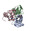
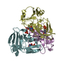
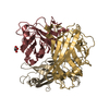
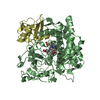
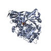
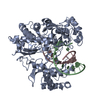
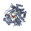
| Title | Crystal structure of ASFV dUTPase | ||||||
 Components Components | E165R | ||||||
 Keywords Keywords | HYDROLASE / dUTPase / ASFV / VIRAL PROTEIN | ||||||
| Function / homology | dUTPase-like / dUTP diphosphatase / dUTPase / dUTP diphosphatase activity / dUTPase, trimeric / dUTPase-like superfamily / Deoxyuridine 5'-triphosphate nucleotidohydrolase Function and homology information Function and homology information | ||||||
| Biological species |   African swine fever virus African swine fever virus | ||||||
| Method |  X-RAY DIFFRACTION / SYNCHROTRON / MOLECULAR REPLACEMENT / Resolution: 2.301 Å X-RAY DIFFRACTION / SYNCHROTRON / MOLECULAR REPLACEMENT / Resolution: 2.301 Å | ||||||
 Authors Authors | Li, C. / Chai, Y. / Song, H. / Qi, J. / Sun, Y. / Gao, G.F. | ||||||
 Citation Citation | Journal: Mbio / Year: 2019 Title: Crystal Structure of African Swine Fever Virus dUTPase Reveals a Potential Drug Target. Authors: Li, C. / Chai, Y. / Song, H. / Weng, C. / Qi, J. / Sun, Y. / Gao, G.F. | ||||||
| History |
|
- Structure visualization
| Structure viewer | Molecule: MolmilJmol/JSmol |
|---|
- Downloads & links
-Download
| PDBx/mmCIF format | 6ky8.cif.gz | 44.2 KB | Display | PDBx/mmCIF format |
|---|---|---|---|---|
| PDB format | pdb6ky8.ent.gz | 29.3 KB | Display | PDB format |
| PDBx/mmJSON format | 6ky8.json.gz | Tree view | PDBx/mmJSON format | |
| Others |  Other downloads Other downloads |
-Validation report
| Arichive directory | https://data.pdbj.org/pub/pdb/validation_reports/ky/6ky8ftp://data.pdbj.org/pub/pdb/validation_reports/ky/6ky8 | HTTPS FTP |
|---|
-Related structure data
| Related structure data | 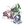 6ky9C  1vyqS S: Starting model for refinement C: citing same article ( |
|---|---|
| Similar structure data |

-Links
 PDBj
PDBj
- Assembly
| Deposited unit | 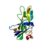
| ||||||||||||
|---|---|---|---|---|---|---|---|---|---|---|---|---|---|
| 1 | 
| ||||||||||||
| Unit cell |
| ||||||||||||
| Components on special symmetry positions |
|
-Components
| #1: Protein | Mass: 19114.328 Da / Num. of mol.: 1 Source method: isolated from a genetically manipulated source Source: (gene. exp.) African swine fever virus / Gene: E165R CDS, E165R, ASFV-Georgia_4-154 / Production host:  |
|---|---|
| #2: Water | ChemComp-HOH /  Mass: 18.015 Da / Num. of mol.: 44 / Source method: isolated from a natural source / Formula: H2O Mass: 18.015 Da / Num. of mol.: 44 / Source method: isolated from a natural source / Formula: H2O |
-Experimental details
-Experiment
| Experiment | Method: X-RAY DIFFRACTION / Number of used crystals: 1 |
|---|
- Sample preparation
| Crystal | Density Matthews: 2.04 Å3/Da / Density % sol: 39.78 % |
|---|---|
| Crystal grow | Temperature: 291 K / Method: vapor diffusion, sitting drop Details: 0.1 M ammonium citrate tribasic pH 7.0, 12% w/v Polyethylene glycol 3,350 |
-Data collection
| Diffraction | Mean temperature: 100 K / Serial crystal experiment: N |
|---|---|
| Diffraction source | Source: SYNCHROTRON / Site: SSRF  / Beamline: BL19U1 / Wavelength: 1.03923 Å / Beamline: BL19U1 / Wavelength: 1.03923 Å |
| Detector | Type: RDI CMOS_8M / Detector: CMOS / Date: Apr 7, 2019 |
| Radiation | Protocol: SINGLE WAVELENGTH / Monochromatic (M) / Laue (L): M / Scattering type: x-ray |
| Radiation wavelength | Wavelength: 1.03923 Å / Relative weight: 1 |
| Reflection | Resolution: 2.3→50 Å / Num. obs: 7082 / % possible obs: 100 % / Redundancy: 38.6 % / CC1/2: 1 / Net I/σ(I): 53.9 |
| Reflection shell | Resolution: 2.3→2.38 Å / Num. unique obs: 690 / CC1/2: 0.999 |
- Processing
| Software |
| ||||||||||||||||||||||||
|---|---|---|---|---|---|---|---|---|---|---|---|---|---|---|---|---|---|---|---|---|---|---|---|---|---|
| Refinement | Method to determine structure: MOLECULAR REPLACEMENT Starting model: 1vyq Resolution: 2.301→39.949 Å / SU ML: 0.19 / Cross valid method: THROUGHOUT / σ(F): 1.36 / Phase error: 27.25
| ||||||||||||||||||||||||
| Solvent computation | Shrinkage radii: 0.9 Å / VDW probe radii: 1.11 Å | ||||||||||||||||||||||||
| Displacement parameters | Biso max: 71.6 Å2 / Biso mean: 30.554 Å2 / Biso min: 11.34 Å2 | ||||||||||||||||||||||||
| Refinement step | Cycle: final / Resolution: 2.301→39.949 Å
| ||||||||||||||||||||||||
| LS refinement shell | Refine-ID: X-RAY DIFFRACTION / Rfactor Rfree error: 0 / % reflection obs: 100 %
|
