 Movie
Movie Controller


+ Open data

- Basic information
| Entry | Database: PDB / ID: 3f0y | ||||||
|---|---|---|---|---|---|---|---|
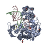
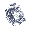
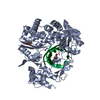
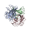
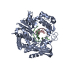
| Title | Crystal structure of the human Adenovirus type 14 fiber knob | ||||||
 Components Components | Fiber protein | ||||||
 Keywords Keywords | VIRAL PROTEIN / adenovirus / Ad14 / CD46 / trimer / fiber / knob | ||||||
| Function / homology |  Function and homology information Function and homology informationadhesion receptor-mediated virion attachment to host cell / viral capsid / cell adhesion / symbiont entry into host cell / host cell nucleus Similarity search - Function | ||||||
| Biological species |  Human adenovirus 14 Human adenovirus 14 | ||||||
| Method |  X-RAY DIFFRACTION / SYNCHROTRON / MOLECULAR REPLACEMENT / Resolution: 1.8 Å X-RAY DIFFRACTION / SYNCHROTRON / MOLECULAR REPLACEMENT / Resolution: 1.8 Å | ||||||
 Authors Authors | Persson, B.D. / Reiter, D.M. / Arnberg, N. / Stehle, T. | ||||||
 Citation Citation | Journal: J.Virol. / Year: 2009 Title: An arginine switch in the species B adenovirus knob determines high-affinity engagement of cellular receptor CD46 Authors: Persson, B.D. / Muller, S. / Reiter, D.M. / Schmitt, B.B. / Marttila, M. / Sumowski, C.V. / Schweizer, S. / Scheu, U. / Ochsenfeld, C. / Arnberg, N. / Stehle, T. | ||||||
| History |
|
- Structure visualization
| Structure viewer | Molecule: MolmilJmol/JSmol |
|---|
- Downloads & links
-Download
| PDBx/mmCIF format | 3f0y.cif.gz | 384.4 KB | Display | PDBx/mmCIF format |
|---|---|---|---|---|
| PDB format | pdb3f0y.ent.gz | 313.4 KB | Display | PDB format |
| PDBx/mmJSON format | 3f0y.json.gz | Tree view | PDBx/mmJSON format | |
| Others |  Other downloads Other downloads |
-Validation report
| Arichive directory | https://data.pdbj.org/pub/pdb/validation_reports/f0/3f0yftp://data.pdbj.org/pub/pdb/validation_reports/f0/3f0y | HTTPS FTP |
|---|
-Related structure data


-Links
 PDBj
PDBj


- Assembly
| Deposited unit | 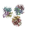
| ||||||||||||||||||
|---|---|---|---|---|---|---|---|---|---|---|---|---|---|---|---|---|---|---|---|
| 1 | 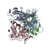
| ||||||||||||||||||
| 2 | 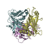
| ||||||||||||||||||
| 3 | 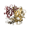
| ||||||||||||||||||
| Unit cell |
| ||||||||||||||||||
| Components on special symmetry positions |
| ||||||||||||||||||
| Noncrystallographic symmetry (NCS) | NCS domain:
|
-Components
| #1: Protein | Mass: 23032.607 Da / Num. of mol.: 9 / Fragment: resdiues 123-325 Source method: isolated from a genetically manipulated source Source: (gene. exp.) Human adenovirus 14 / Strain: De Wit / Gene: Fiber, L5 / Plasmid: pET15b / Production host:  #2: Chemical | ChemComp-GOL /   Mass: 92.094 Da / Num. of mol.: 4 / Source method: obtained synthetically / Formula: C3H8O3 Mass: 92.094 Da / Num. of mol.: 4 / Source method: obtained synthetically / Formula: C3H8O3#3: Chemical |   Mass: 69.085 Da / Num. of mol.: 3 / Source method: obtained synthetically / Formula: C3H5N2 Mass: 69.085 Da / Num. of mol.: 3 / Source method: obtained synthetically / Formula: C3H5N2#4: Water | ChemComp-HOH / |  Mass: 18.015 Da / Num. of mol.: 1803 / Source method: isolated from a natural source / Formula: H2O Mass: 18.015 Da / Num. of mol.: 1803 / Source method: isolated from a natural source / Formula: H2O |
|---|
-Experimental details
-Experiment
| Experiment | Method: X-RAY DIFFRACTION / Number of used crystals: 1 |
|---|
- Sample preparation
| Crystal | Density Matthews: 2.46 Å3/Da / Density % sol: 50.01 % |
|---|---|
| Crystal grow | Temperature: 293.15 K / Method: vapor diffusion, sitting drop / pH: 9 Details: 20% (w/v) PEG 8000, 0.1M CHES, 200mM NaCl, pH 9.0, VAPOR DIFFUSION, SITTING DROP, temperature 293.15K |
-Data collection
| Diffraction | Mean temperature: 93.15 K |
|---|---|
| Diffraction source | Source: SYNCHROTRON / Site: SLS  / Beamline: X06SA / Wavelength: 1 Å / Beamline: X06SA / Wavelength: 1 Å |
| Detector | Type: PSI PILATUS 6M / Detector: PIXEL / Date: Nov 11, 2007 |
| Radiation | Monochromator: Undulator / Protocol: SINGLE WAVELENGTH / Monochromatic (M) / Laue (L): M / Scattering type: x-ray |
| Radiation wavelength | Wavelength: 1 Å / Relative weight: 1 |
| Reflection | Resolution: 1.8→47.4 Å / Num. all: 187138 / Num. obs: 185386 / % possible obs: 99.1 % / Observed criterion σ(F): 0 / Observed criterion σ(I): -3 / Rmerge(I) obs: 0.078 |
| Reflection shell | Resolution: 1.8→1.9 Å / % possible all: 94.4 |
- Processing
| Software |
| |||||||||||||||||||||||||||||||||||||||||||||||||||||||||||||||||||||||||||||||||||||||||||||||||||||||||||||||||||||||||||||
|---|---|---|---|---|---|---|---|---|---|---|---|---|---|---|---|---|---|---|---|---|---|---|---|---|---|---|---|---|---|---|---|---|---|---|---|---|---|---|---|---|---|---|---|---|---|---|---|---|---|---|---|---|---|---|---|---|---|---|---|---|---|---|---|---|---|---|---|---|---|---|---|---|---|---|---|---|---|---|---|---|---|---|---|---|---|---|---|---|---|---|---|---|---|---|---|---|---|---|---|---|---|---|---|---|---|---|---|---|---|---|---|---|---|---|---|---|---|---|---|---|---|---|---|---|---|---|
| Refinement | Method to determine structure: MOLECULAR REPLACEMENT Starting model: Human Adenovirus type 11 fiber knob Resolution: 1.8→47.4 Å / Cor.coef. Fo:Fc: 0.95 / Cor.coef. Fo:Fc free: 0.938 / SU B: 2.745 / SU ML: 0.085 / Cross valid method: THROUGHOUT / σ(F): 0 / σ(I): 0 / ESU R: 0.129 / ESU R Free: 0.118 / Stereochemistry target values: MAXIMUM LIKELIHOOD / Details: HYDROGENS HAVE BEEN ADDED IN THE RIDING POSITIONS
| |||||||||||||||||||||||||||||||||||||||||||||||||||||||||||||||||||||||||||||||||||||||||||||||||||||||||||||||||||||||||||||
| Solvent computation | Ion probe radii: 0.8 Å / Shrinkage radii: 0.8 Å / VDW probe radii: 1.2 Å / Solvent model: MASK | |||||||||||||||||||||||||||||||||||||||||||||||||||||||||||||||||||||||||||||||||||||||||||||||||||||||||||||||||||||||||||||
| Displacement parameters | Biso mean: 14.92 Å2
| |||||||||||||||||||||||||||||||||||||||||||||||||||||||||||||||||||||||||||||||||||||||||||||||||||||||||||||||||||||||||||||
| Refinement step | Cycle: LAST / Resolution: 1.8→47.4 Å
| |||||||||||||||||||||||||||||||||||||||||||||||||||||||||||||||||||||||||||||||||||||||||||||||||||||||||||||||||||||||||||||
| Refine LS restraints |
| |||||||||||||||||||||||||||||||||||||||||||||||||||||||||||||||||||||||||||||||||||||||||||||||||||||||||||||||||||||||||||||
| Refine LS restraints NCS | Dom-ID: 1 / Refine-ID: X-RAY DIFFRACTION
| |||||||||||||||||||||||||||||||||||||||||||||||||||||||||||||||||||||||||||||||||||||||||||||||||||||||||||||||||||||||||||||
| LS refinement shell | Resolution: 1.8→1.848 Å / Total num. of bins used: 20
|
