 Movie
Movie Controller


+ Open data

- Basic information
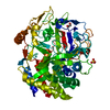
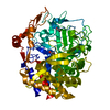
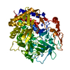
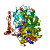
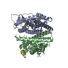
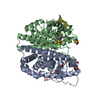
| Entry | Database: PDB / ID: 4jjj | |||||||||
|---|---|---|---|---|---|---|---|---|---|---|
| Title | The structure of T. fusca GH48 D224N mutant | |||||||||
 Components Components | Cellulose 1,4-beta-cellobiosidase | |||||||||
 Keywords Keywords | HYDROLASE / GH48 / (a/a)6 / cellobiohydrolase | |||||||||
| Function / homology |  Function and homology information Function and homology informationcellulose 1,4-beta-cellobiosidase (non-reducing end) / cellulose 1,4-beta-cellobiosidase activity / cellulase activity / polysaccharide binding / cellulose catabolic process / metal ion binding Similarity search - Function | |||||||||
| Biological species |   Thermobifida fusca (bacteria) Thermobifida fusca (bacteria) | |||||||||
| Method |  X-RAY DIFFRACTION / MOLECULAR REPLACEMENT / Resolution: 1.6 Å X-RAY DIFFRACTION / MOLECULAR REPLACEMENT / Resolution: 1.6 Å | |||||||||
 Authors Authors | Alahuhta, P.M. / Lunin, V.V. | |||||||||
 Citation Citation | Journal: Biotechnol.Bioeng. / Year: 2014 Title: Cel48A from Thermobifida fusca: structure and site directed mutagenesis of key residues. Authors: Kostylev, M. / Alahuhta, M. / Chen, M. / Brunecky, R. / Himmel, M.E. / Lunin, V.V. / Brady, J. / Wilson, D.B. | |||||||||
| History |
|
- Structure visualization
| Structure viewer | Molecule: MolmilJmol/JSmol |
|---|
- Downloads & links
-Download
| PDBx/mmCIF format | 4jjj.cif.gz | 189.1 KB | Display | PDBx/mmCIF format |
|---|---|---|---|---|
| PDB format | pdb4jjj.ent.gz | 144.4 KB | Display | PDB format |
| PDBx/mmJSON format | 4jjj.json.gz | Tree view | PDBx/mmJSON format | |
| Others |  Other downloads Other downloads |
-Validation report
| Arichive directory | https://data.pdbj.org/pub/pdb/validation_reports/jj/4jjjftp://data.pdbj.org/pub/pdb/validation_reports/jj/4jjj | HTTPS FTP |
|---|
-Related structure data
| Related structure data |  4fusS S: Starting model for refinement |
|---|---|
| Similar structure data |


-Links
 PDBj
PDBj

- Assembly
| Deposited unit | 
| ||||||||
|---|---|---|---|---|---|---|---|---|---|
| 1 |
| ||||||||
| Unit cell |
|
-Components
-Protein , 1 types, 1 molecules A
| #1: Protein | Mass: 71759.320 Da / Num. of mol.: 1 / Fragment: UNP residues 343-984 / Mutation: D224N Source method: isolated from a genetically manipulated source Source: (gene. exp.) Thermobifida fusca (bacteria) / Gene: Tfu_1959 / Production host: References: UniProt: Q47NH7, cellulose 1,4-beta-cellobiosidase (non-reducing end) |
|---|
-Sugars , 3 types, 3 molecules
| #2: Polysaccharide | beta-D-glucopyranose-(1-4)-beta-D-glucopyranose / beta-cellobiose  Source method: isolated from a genetically manipulated source Details: oligosaccharide / References: beta-cellobiose |
|---|---|
| #3: Polysaccharide | beta-D-glucopyranose-(1-4)-alpha-D-glucopyranose / alpha-cellobiose  Source method: isolated from a genetically manipulated source Details: oligosaccharide / References: alpha-cellobiose |
| #4: Polysaccharide | beta-D-glucopyranose-(1-4)-beta-D-glucopyranose-(1-4)-beta-D-glucopyranose-(1-4)-beta-D- ...beta-D-glucopyranose-(1-4)-beta-D-glucopyranose-(1-4)-beta-D-glucopyranose-(1-4)-beta-D-glucopyranose-(1-4)-beta-D-glucopyranose-(1-4)-beta-D-glucopyranose / beta-cellohexaose  Source method: isolated from a genetically manipulated source Details: oligosaccharide / References: beta-cellohexaose |
-Non-polymers , 7 types, 1171 molecules 


| #5: Chemical | ChemComp-CA /  Mass: 40.078 Da / Num. of mol.: 4 / Source method: obtained synthetically / Formula: Ca Mass: 40.078 Da / Num. of mol.: 4 / Source method: obtained synthetically / Formula: Ca#6: Chemical |  Mass: 22.990 Da / Num. of mol.: 3 / Source method: obtained synthetically / Formula: Na Mass: 22.990 Da / Num. of mol.: 3 / Source method: obtained synthetically / Formula: Na#7: Chemical | ChemComp-FE /  Mass: 55.845 Da / Num. of mol.: 4 / Source method: obtained synthetically / Formula: Fe Mass: 55.845 Da / Num. of mol.: 4 / Source method: obtained synthetically / Formula: Fe#8: Chemical | ChemComp-ZN /  Mass: 65.409 Da / Num. of mol.: 21 / Source method: obtained synthetically / Formula: Zn Mass: 65.409 Da / Num. of mol.: 21 / Source method: obtained synthetically / Formula: Zn#9: Chemical |  Mass: 59.044 Da / Num. of mol.: 2 / Source method: obtained synthetically / Formula: C2H3O2 Mass: 59.044 Da / Num. of mol.: 2 / Source method: obtained synthetically / Formula: C2H3O2#10: Chemical | ChemComp-EDO / |  Mass: 62.068 Da / Num. of mol.: 1 / Source method: obtained synthetically / Formula: C2H6O2 Mass: 62.068 Da / Num. of mol.: 1 / Source method: obtained synthetically / Formula: C2H6O2#11: Water | ChemComp-HOH / | Mass: 18.015 Da / Num. of mol.: 1136 / Source method: isolated from a natural source / Formula: H2O |
|---|
-Details
| Has protein modification | Y |
|---|
-Experimental details
-Experiment
| Experiment | Method: X-RAY DIFFRACTION / Number of used crystals: 1 |
|---|
- Sample preparation
| Crystal | Density Matthews: 2.13 Å3/Da / Density % sol: 42.19 % |
|---|---|
| Crystal grow | Temperature: 294 K / Method: vapor diffusion, sitting drop / pH: 6.5 Details: 0.1 M sodium cacodylate trihydrate pH 6.5, 18% (w/v) PEG 8000 and 20 mM zinc acetate dihydrate, VAPOR DIFFUSION, SITTING DROP, temperature 294K |
-Data collection
| Diffraction | Mean temperature: 100 K |
|---|---|
| Diffraction source | Source: ROTATING ANODE / Type: BRUKER AXS MICROSTAR / Wavelength: 1.5418 Å |
| Detector | Type: Bruker Platinum 135 / Detector: CCD / Date: Apr 30, 2012 |
| Radiation | Monochromator: HELIOS MIRRORS / Protocol: SINGLE WAVELENGTH / Monochromatic (M) / Laue (L): M / Scattering type: x-ray |
| Radiation wavelength | Wavelength: 1.5418 Å / Relative weight: 1 |
| Reflection | Resolution: 1.6→25 Å / Num. all: 78727 / Num. obs: 78727 / % possible obs: 99.4 % / Redundancy: 7.93 % / Rsym value: 0.0886 / Net I/σ(I): 15.1 |
| Reflection shell | Resolution: 1.6→1.7 Å / Redundancy: 3.32 % / Mean I/σ(I) obs: 3.81 / Num. unique all: 12722 / Rsym value: 0.2506 / % possible all: 93.9 |
- Processing
| Software |
| ||||||||||||||||||||||||||||||||||||||||||||||||||||||||||||||||||||||||||||||||||||||||||||||||||||||||||||||||||||||||||||||||||||||||||||||||||||||||||||||||||||||||||
|---|---|---|---|---|---|---|---|---|---|---|---|---|---|---|---|---|---|---|---|---|---|---|---|---|---|---|---|---|---|---|---|---|---|---|---|---|---|---|---|---|---|---|---|---|---|---|---|---|---|---|---|---|---|---|---|---|---|---|---|---|---|---|---|---|---|---|---|---|---|---|---|---|---|---|---|---|---|---|---|---|---|---|---|---|---|---|---|---|---|---|---|---|---|---|---|---|---|---|---|---|---|---|---|---|---|---|---|---|---|---|---|---|---|---|---|---|---|---|---|---|---|---|---|---|---|---|---|---|---|---|---|---|---|---|---|---|---|---|---|---|---|---|---|---|---|---|---|---|---|---|---|---|---|---|---|---|---|---|---|---|---|---|---|---|---|---|---|---|---|---|---|
| Refinement | Method to determine structure: MOLECULAR REPLACEMENT Starting model: 4FUS Resolution: 1.6→25 Å / Cor.coef. Fo:Fc: 0.976 / Cor.coef. Fo:Fc free: 0.958 / SU B: 1.343 / SU ML: 0.048 / Cross valid method: THROUGHOUT / ESU R: 0.075 / ESU R Free: 0.08 / Stereochemistry target values: MAXIMUM LIKELIHOOD / Details: HYDROGENS HAVE BEEN ADDED IN THE RIDING POSITIONS
| ||||||||||||||||||||||||||||||||||||||||||||||||||||||||||||||||||||||||||||||||||||||||||||||||||||||||||||||||||||||||||||||||||||||||||||||||||||||||||||||||||||||||||
| Solvent computation | Ion probe radii: 0.8 Å / Shrinkage radii: 0.8 Å / VDW probe radii: 1.2 Å / Solvent model: MASK | ||||||||||||||||||||||||||||||||||||||||||||||||||||||||||||||||||||||||||||||||||||||||||||||||||||||||||||||||||||||||||||||||||||||||||||||||||||||||||||||||||||||||||
| Displacement parameters | Biso mean: 11.472 Å2
| ||||||||||||||||||||||||||||||||||||||||||||||||||||||||||||||||||||||||||||||||||||||||||||||||||||||||||||||||||||||||||||||||||||||||||||||||||||||||||||||||||||||||||
| Refinement step | Cycle: LAST / Resolution: 1.6→25 Å
| ||||||||||||||||||||||||||||||||||||||||||||||||||||||||||||||||||||||||||||||||||||||||||||||||||||||||||||||||||||||||||||||||||||||||||||||||||||||||||||||||||||||||||
| Refine LS restraints |
| ||||||||||||||||||||||||||||||||||||||||||||||||||||||||||||||||||||||||||||||||||||||||||||||||||||||||||||||||||||||||||||||||||||||||||||||||||||||||||||||||||||||||||
| LS refinement shell | Resolution: 1.6→1.642 Å / Total num. of bins used: 20
|
