 Movie
Movie Controller


+ Open data

- Basic information
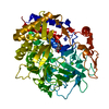
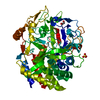
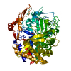
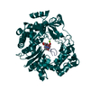
| Entry | Database: PDB / ID: 1g9g | ||||||
|---|---|---|---|---|---|---|---|
| Title | XTAL-STRUCTURE OF THE FREE NATIVE CELLULASE CEL48F | ||||||
 Components Components | CELLULASE CEL48F | ||||||
 Keywords Keywords | HYDROLASE / Cellulase / processive-endo | ||||||
| Function / homology |  Function and homology information Function and homology information | ||||||
| Biological species |  Clostridium cellulolyticum (bacteria) Clostridium cellulolyticum (bacteria) | ||||||
| Method |  X-RAY DIFFRACTION / SYNCHROTRON / MOLECULAR REPLACEMENT / Resolution: 1.9 Å X-RAY DIFFRACTION / SYNCHROTRON / MOLECULAR REPLACEMENT / Resolution: 1.9 Å | ||||||
 Authors Authors | Parsiegla, G. / Tardif, C. / Belaich, J.P. / Driguez, H. / Haser, R. | ||||||
 Citation Citation | Journal: J.Mol.Biol. / Year: 2008 Title: Structures of mutants of cellulase Cel48F of Clostridium cellulolyticum in complex with long hemithiocellooligosaccharides give rise to a new view of the substrate pathway during processive action Authors: Parsiegla, G. / Reverbel, C. / Tardif, C. / Driguez, H. / Haser, R. #1: Journal: Biochemistry / Year: 2000Title: Crystal structures of the cellulase Cel48F in complex with inhibitors and substrates give insights into its processive action. Authors: Parsiegla, G. / Reverbel-Leroy, C. / Tardif, C. / Belaich, J.P. / Driguez, H. / Haser, R. #2: Journal: Embo J. / Year: 1998Title: The crystal structure of the processive endocellulase CelF of Clostridium cellulolyticum in complex with a thioligosaccharide inhibitor at 2.0 a resolution. Authors: Parsiegla, G. / Juy, M. / Reverbel-Leroy, C. / Tardif, C. / Belaich, J.P. / Driguez, H. / Haser, R. | ||||||
| History |
|
- Structure visualization
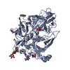
| Structure viewer | Molecule: MolmilJmol/JSmol |
|---|
- Downloads & links
-Download
| PDBx/mmCIF format | 1g9g.cif.gz | 147.7 KB | Display | PDBx/mmCIF format |
|---|---|---|---|---|
| PDB format | pdb1g9g.ent.gz | 113.8 KB | Display | PDB format |
| PDBx/mmJSON format | 1g9g.json.gz | Tree view | PDBx/mmJSON format | |
| Others |  Other downloads Other downloads |
-Validation report
| Arichive directory | https://data.pdbj.org/pub/pdb/validation_reports/g9/1g9gftp://data.pdbj.org/pub/pdb/validation_reports/g9/1g9g | HTTPS FTP |
|---|
-Related structure data
| Related structure data | 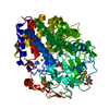 1g9jC 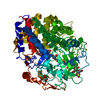 2qnoC 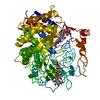 1fceS C: citing same article ( S: Starting model for refinement |
|---|---|
| Similar structure data |


-Links
 PDBj
PDBj
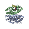
- Assembly
| Deposited unit | 
| ||||||||
|---|---|---|---|---|---|---|---|---|---|
| 1 |
| ||||||||
| Unit cell |
|
-Components
| #1: Protein | Mass: 70869.875 Da / Num. of mol.: 1 / Fragment: CATALYTIC MODULE Source method: isolated from a genetically manipulated source Source: (gene. exp.) Clostridium cellulolyticum (bacteria) / Plasmid: PETFC / Species (production host): Escherichia coli / Production host: |
|---|---|
| #2: Chemical | ChemComp-CA /   Mass: 40.078 Da / Num. of mol.: 1 / Source method: obtained synthetically / Formula: Ca Mass: 40.078 Da / Num. of mol.: 1 / Source method: obtained synthetically / Formula: Ca |
| #3: Chemical | ChemComp-MG /   Mass: 24.305 Da / Num. of mol.: 1 / Source method: obtained synthetically / Formula: Mg Mass: 24.305 Da / Num. of mol.: 1 / Source method: obtained synthetically / Formula: Mg |
| #4: Water | ChemComp-HOH /  Mass: 18.015 Da / Num. of mol.: 397 / Source method: isolated from a natural source / Formula: H2O Mass: 18.015 Da / Num. of mol.: 397 / Source method: isolated from a natural source / Formula: H2O |
-Experimental details
-Experiment
| Experiment | Method: X-RAY DIFFRACTION / Number of used crystals: 1 |
|---|
- Sample preparation
| Crystal | Density Matthews: 2.25 Å3/Da / Density % sol: 45.23 % | |||||||||||||||||||||||||||||||||||
|---|---|---|---|---|---|---|---|---|---|---|---|---|---|---|---|---|---|---|---|---|---|---|---|---|---|---|---|---|---|---|---|---|---|---|---|---|
| Crystal grow | Temperature: 290 K / Method: vapor diffusion, hanging drop / pH: 7.5 Details: HEPES, PEG 4000, MgCl, Glucose, pH 7.5, VAPOR DIFFUSION, HANGING DROP, temperature 290K | |||||||||||||||||||||||||||||||||||
| Crystal grow | *PLUS Temperature: 293 K / Method: vapor diffusion, hanging dropDetails: Reverbel-Leroy, C., (1997) Acta Crystallogr., D.54, 114. | |||||||||||||||||||||||||||||||||||
| Components of the solutions | *PLUS
|
-Data collection
| Diffraction | Mean temperature: 290 K |
|---|---|
| Diffraction source | Source: SYNCHROTRON / Site: ESRF  / Beamline: BM30A / Wavelength: 0.9796 Å / Beamline: BM30A / Wavelength: 0.9796 Å |
| Detector | Type: MARRESEARCH / Detector: IMAGE PLATE / Date: Aug 30, 1999 |
| Radiation | Monochromator: Synchrotron / Protocol: SINGLE WAVELENGTH / Monochromatic (M) / Laue (L): M / Scattering type: x-ray |
| Radiation wavelength | Wavelength: 0.9796 Å / Relative weight: 1 |
| Reflection | Resolution: 1.85→40 Å / Num. all: 54275 / Num. obs: 49813 / % possible obs: 98.2 % / Observed criterion σ(I): 2 / Redundancy: 3.8 % / Biso Wilson estimate: 12.7 Å2 / Rmerge(I) obs: 0.091 / Net I/σ(I): 6.8 |
| Reflection shell | Resolution: 1.85→1.9 Å / Redundancy: 2.3 % / Rmerge(I) obs: 0.354 / Mean I/σ(I) obs: 1.9 / Num. unique all: 2584 / % possible all: 88.9 |
| Reflection shell | *PLUS % possible obs: 88.9 % |
- Processing
| Software |
| ||||||||||||||||||||||||||||||||||||||||||||||||||||||||||||||||||||||||||||||||
|---|---|---|---|---|---|---|---|---|---|---|---|---|---|---|---|---|---|---|---|---|---|---|---|---|---|---|---|---|---|---|---|---|---|---|---|---|---|---|---|---|---|---|---|---|---|---|---|---|---|---|---|---|---|---|---|---|---|---|---|---|---|---|---|---|---|---|---|---|---|---|---|---|---|---|---|---|---|---|---|---|---|
| Refinement | Method to determine structure: MOLECULAR REPLACEMENT Starting model: PDB ENTRY 1FCE Resolution: 1.9→38.59 Å / Rfactor Rfree error: 0.004 / Data cutoff high absF: 2238873.49 / Data cutoff low absF: 0 / Isotropic thermal model: RESTRAINED / Cross valid method: THROUGHOUT / σ(F): 0 / Stereochemistry target values: Engh & Huber
| ||||||||||||||||||||||||||||||||||||||||||||||||||||||||||||||||||||||||||||||||
| Solvent computation | Solvent model: FLAT MODEL / Bsol: 36.74 Å2 / ksol: 0.3 e/Å3 | ||||||||||||||||||||||||||||||||||||||||||||||||||||||||||||||||||||||||||||||||
| Displacement parameters | Biso mean: 19.4 Å2
| ||||||||||||||||||||||||||||||||||||||||||||||||||||||||||||||||||||||||||||||||
| Refine analyze |
| ||||||||||||||||||||||||||||||||||||||||||||||||||||||||||||||||||||||||||||||||
| Refinement step | Cycle: LAST / Resolution: 1.9→38.59 Å
| ||||||||||||||||||||||||||||||||||||||||||||||||||||||||||||||||||||||||||||||||
| Refine LS restraints |
| ||||||||||||||||||||||||||||||||||||||||||||||||||||||||||||||||||||||||||||||||
| LS refinement shell | Resolution: 1.9→2.02 Å / Rfactor Rfree error: 0.013 / Total num. of bins used: 6
| ||||||||||||||||||||||||||||||||||||||||||||||||||||||||||||||||||||||||||||||||
| Xplor file |
| ||||||||||||||||||||||||||||||||||||||||||||||||||||||||||||||||||||||||||||||||
| Refinement | *PLUS Highest resolution: 1.85 Å / Lowest resolution: 40 Å | ||||||||||||||||||||||||||||||||||||||||||||||||||||||||||||||||||||||||||||||||
| Solvent computation | *PLUS | ||||||||||||||||||||||||||||||||||||||||||||||||||||||||||||||||||||||||||||||||
| Displacement parameters | *PLUS | ||||||||||||||||||||||||||||||||||||||||||||||||||||||||||||||||||||||||||||||||
| Refine LS restraints | *PLUS
|
