Movie
Movie Controller

[English] 日本語
 Yorodumi
- PDB-6kmw: Structure of PSI from H. hongdechloris grown under white light co... -
+ Open data

- Basic information
| Entry | Database: PDB / ID: 6kmw | |||||||||
|---|---|---|---|---|---|---|---|---|---|---|
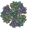
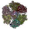
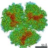
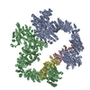
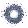
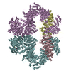
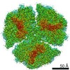
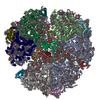
| Title | Structure of PSI from H. hongdechloris grown under white light condition | |||||||||
 Components Components |
| |||||||||
 Keywords Keywords | ELECTRON TRANSPORT / Photosystem I | |||||||||
| Function / homology |  Function and homology information Function and homology informationphotosystem I reaction center / photosystem I / photosynthetic electron transport in photosystem I / photosystem I / plasma membrane-derived thylakoid membrane / chlorophyll binding / photosynthesis / endomembrane system / 4 iron, 4 sulfur cluster binding / electron transfer activity ...photosystem I reaction center / photosystem I / photosynthetic electron transport in photosystem I / photosystem I / plasma membrane-derived thylakoid membrane / chlorophyll binding / photosynthesis / endomembrane system / 4 iron, 4 sulfur cluster binding / electron transfer activity / oxidoreductase activity / magnesium ion binding / metal ion binding Similarity search - Function | |||||||||
| Biological species |  Halomicronema hongdechloris C2206 (bacteria) Halomicronema hongdechloris C2206 (bacteria) | |||||||||
| Method | ELECTRON MICROSCOPY / single particle reconstruction / cryo EM / Resolution: 2.35 Å | |||||||||
 Authors Authors | Kato, K. / Nagao, R. / Shen, J.R. / Miyazaki, N. / Akita, F. | |||||||||
 Citation Citation | Journal: Nat Commun / Year: 2020 Title: Structural basis for the adaptation and function of chlorophyll f in photosystem I. Authors: Koji Kato / Toshiyuki Shinoda / Ryo Nagao / Seiji Akimoto / Takehiro Suzuki / Naoshi Dohmae / Min Chen / Suleyman I Allakhverdiev / Jian-Ren Shen / Fusamichi Akita / Naoyuki Miyazaki / Tatsuya Tomo /    Abstract: Chlorophylls (Chl) play pivotal roles in energy capture, transfer and charge separation in photosynthesis. Among Chls functioning in oxygenic photosynthesis, Chl f is the most red-shifted type first ...Chlorophylls (Chl) play pivotal roles in energy capture, transfer and charge separation in photosynthesis. Among Chls functioning in oxygenic photosynthesis, Chl f is the most red-shifted type first found in a cyanobacterium Halomicronema hongdechloris. The location and function of Chl f in photosystems are not clear. Here we analyzed the high-resolution structures of photosystem I (PSI) core from H. hongdechloris grown under white or far-red light by cryo-electron microscopy. The structure showed that, far-red PSI binds 83 Chl a and 7 Chl f, and Chl f are associated at the periphery of PSI but not in the electron transfer chain. The appearance of Chl f is well correlated with the expression of PSI genes induced under far-red light. These results indicate that Chl f functions to harvest the far-red light and enhance uphill energy transfer, and changes in the gene sequences are essential for the binding of Chl f. | |||||||||
| History |
|
- Structure visualization
| Movie |
Movie viewer |
|---|---|
| Structure viewer | Molecule: MolmilJmol/JSmol |
- Downloads & links
-Download
| PDBx/mmCIF format | 6kmw.cif.gz | 1.4 MB | Display | PDBx/mmCIF format |
|---|---|---|---|---|
| PDB format | pdb6kmw.ent.gz | Display | PDB format | |
| PDBx/mmJSON format | 6kmw.json.gz | Tree view | PDBx/mmJSON format | |
| Others |  Other downloads Other downloads |
-Validation report
| Arichive directory | https://data.pdbj.org/pub/pdb/validation_reports/km/6kmwftp://data.pdbj.org/pub/pdb/validation_reports/km/6kmw | HTTPS FTP |
|---|
-Related structure data
| Related structure data | 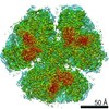 0726MC  0727C 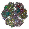 6kmxC M: map data used to model this data C: citing same article ( |
|---|---|
| Similar structure data |

-Links
 PDBj
PDBj


- Assembly
| Deposited unit | 
|
|---|---|
| 1 |
|
-Components
-Photosystem I P700 chlorophyll a apoprotein ... , 2 types, 6 molecules aAbAcAaBbBcB
| #1: Protein | Mass: 84273.633 Da / Num. of mol.: 3 / Source method: isolated from a natural source Source: (natural) Halomicronema hongdechloris C2206 (bacteria)References: UniProt: A0A1Z3HRW4, photosystem I #2: Protein | Mass: 83080.469 Da / Num. of mol.: 3 / Source method: isolated from a natural source Source: (natural) Halomicronema hongdechloris C2206 (bacteria)References: UniProt: A0A1Z3HRY4, photosystem I |
|---|
-Photosystem I reaction center subunit ... , 5 types, 15 molecules aDbDcDaEbEcEaIbIcIaLbLcLaMbMcM
| #4: Protein | Mass: 15723.800 Da / Num. of mol.: 3 / Source method: isolated from a natural source Source: (natural) Halomicronema hongdechloris C2206 (bacteria)References: UniProt: A0A1Z3HHI7, photosystem I #5: Protein | Mass: 7907.028 Da / Num. of mol.: 3 / Source method: isolated from a natural source Source: (natural) Halomicronema hongdechloris C2206 (bacteria)References: UniProt: A0A1Z3HI16 #6: Protein/peptide | Mass: 4318.124 Da / Num. of mol.: 3 / Source method: isolated from a natural source Source: (natural) Halomicronema hongdechloris C2206 (bacteria)References: UniProt: A0A1Z3HSF0, photosystem I #7: Protein | Mass: 16871.471 Da / Num. of mol.: 3 / Source method: isolated from a natural source Source: (natural) Halomicronema hongdechloris C2206 (bacteria)References: UniProt: A0A1Z3HS05, photosystem I #8: Protein/peptide | Mass: 3440.141 Da / Num. of mol.: 3 / Source method: isolated from a natural source Source: (natural) Halomicronema hongdechloris C2206 (bacteria) |
|---|
-Protein / Sugars , 2 types, 6 molecules aCbCcC
| #15: Sugar |  Type: D-saccharide / Mass: 510.615 Da / Num. of mol.: 3 / Source method: obtained synthetically / Formula: C24H46O11 / Comment: detergent*YM Type: D-saccharide / Mass: 510.615 Da / Num. of mol.: 3 / Source method: obtained synthetically / Formula: C24H46O11 / Comment: detergent*YM#3: Protein | Mass: 8782.184 Da / Num. of mol.: 3 / Source method: isolated from a natural source Source: (natural) Halomicronema hongdechloris C2206 (bacteria)References: UniProt: A0A1Z3HPE3, photosystem I |
|---|
-Non-polymers , 10 types, 990 molecules 


| #9: Chemical |  Mass: 893.489 Da / Num. of mol.: 3 / Source method: obtained synthetically / Formula: C55H72MgN4O5 Mass: 893.489 Da / Num. of mol.: 3 / Source method: obtained synthetically / Formula: C55H72MgN4O5#10: Chemical | ChemComp-CLA /  Mass: 893.489 Da / Num. of mol.: 267 / Source method: obtained synthetically / Formula: C55H72MgN4O5 Mass: 893.489 Da / Num. of mol.: 267 / Source method: obtained synthetically / Formula: C55H72MgN4O5#11: Chemical | ChemComp-PQN /  Mass: 450.696 Da / Num. of mol.: 6 / Source method: obtained synthetically / Formula: C31H46O2 Mass: 450.696 Da / Num. of mol.: 6 / Source method: obtained synthetically / Formula: C31H46O2#12: Chemical | ChemComp-SF4 /  Mass: 351.640 Da / Num. of mol.: 9 / Source method: obtained synthetically / Formula: Fe4S4 Mass: 351.640 Da / Num. of mol.: 9 / Source method: obtained synthetically / Formula: Fe4S4#13: Chemical | ChemComp-BCR /  Mass: 536.873 Da / Num. of mol.: 51 / Source method: obtained synthetically / Formula: C40H56 Mass: 536.873 Da / Num. of mol.: 51 / Source method: obtained synthetically / Formula: C40H56#14: Chemical | ChemComp-LHG /  Mass: 722.970 Da / Num. of mol.: 6 / Source method: obtained synthetically / Formula: C38H75O10P / Comment: phospholipid*YM Mass: 722.970 Da / Num. of mol.: 6 / Source method: obtained synthetically / Formula: C38H75O10P / Comment: phospholipid*YM#16: Chemical |  Mass: 787.158 Da / Num. of mol.: 3 / Source method: obtained synthetically / Formula: C45H86O10 Mass: 787.158 Da / Num. of mol.: 3 / Source method: obtained synthetically / Formula: C45H86O10#17: Chemical | ChemComp-UNL / Num. of mol.: 15 / Source method: obtained synthetically #18: Chemical |  Mass: 40.078 Da / Num. of mol.: 3 / Source method: obtained synthetically / Formula: Ca Mass: 40.078 Da / Num. of mol.: 3 / Source method: obtained synthetically / Formula: Ca#19: Water | ChemComp-HOH / | Mass: 18.015 Da / Num. of mol.: 627 / Source method: isolated from a natural source / Formula: H2O |
|---|
-Details
| Has ligand of interest | N |
|---|
-Experimental details
-Experiment
| Experiment | Method: ELECTRON MICROSCOPY |
|---|---|
| EM experiment | Aggregation state: PARTICLE / 3D reconstruction method: single particle reconstruction |
- Sample preparation
| Component | Name: white PSI / Type: COMPLEX / Entity ID: #1-#8 / Source: NATURAL | |||||||||||||||
|---|---|---|---|---|---|---|---|---|---|---|---|---|---|---|---|---|
| Molecular weight | Experimental value: NO | |||||||||||||||
| Source (natural) | Organism: Halomicronema hongdechloris C2206 (bacteria) | |||||||||||||||
| Buffer solution | pH: 6.5 | |||||||||||||||
| Buffer component |
| |||||||||||||||
| Specimen | Conc.: 0.032 mg/ml / Embedding applied: NO / Shadowing applied: NO / Staining applied: NO / Vitrification applied: YES | |||||||||||||||
| Specimen support | Grid material: COPPER / Grid mesh size: 200 divisions/in. / Grid type: Quantifoil R1.2/1.3 | |||||||||||||||
| Vitrification | Instrument: FEI VITROBOT MARK IV / Cryogen name: ETHANE / Humidity: 100 % / Chamber temperature: 277 K |
- Electron microscopy imaging
| Experimental equipment |  Model: Titan Krios / Image courtesy: FEI Company |
|---|---|
| Microscopy | Model: FEI TITAN KRIOS |
| Electron gun | Electron source:  FIELD EMISSION GUN / Accelerating voltage: 300 kV / Illumination mode: FLOOD BEAM FIELD EMISSION GUN / Accelerating voltage: 300 kV / Illumination mode: FLOOD BEAM |
| Electron lens | Mode: BRIGHT FIELD |
| Image recording | Electron dose: 47 e/Å2 / Detector mode: COUNTING / Film or detector model: FEI FALCON III (4k x 4k) |
- Processing
| EM software |
| ||||||||||||||||
|---|---|---|---|---|---|---|---|---|---|---|---|---|---|---|---|---|---|
| CTF correction | Type: PHASE FLIPPING AND AMPLITUDE CORRECTION | ||||||||||||||||
| Symmetry | Point symmetry: C3 (3 fold cyclic) | ||||||||||||||||
| 3D reconstruction | Resolution: 2.35 Å / Resolution method: FSC 0.143 CUT-OFF / Num. of particles: 546366 / Symmetry type: POINT | ||||||||||||||||
| Atomic model building | Protocol: FLEXIBLE FIT / Space: REAL / Target criteria: Correlation coefficient | ||||||||||||||||
| Atomic model building | PDB-ID: 1JB0 Accession code: 1JB0 / Source name: PDB / Type: experimental model |