 Movie
Movie Controller


+ Open data

- Basic information
| Entry | Database: PDB / ID: 6z2w | ||||||
|---|---|---|---|---|---|---|---|
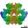
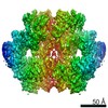
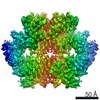
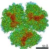
| Title | Mec1-Ddc2 (F2244L mutant) in complex with Mg AMP-PNP | ||||||
 Components Components |
| ||||||
 Keywords Keywords | HYDROLASE / serine/threonine protein kinase / complex / DNA damage response / checkpoint control | ||||||
| Function / homology |  Function and homology information Function and homology informationATR-ATRIP complex / positive regulation of DNA-templated DNA replication / regulation of double-strand break repair / telomere maintenance via recombination / nucleobase-containing compound metabolic process / reciprocal meiotic recombination / nuclear chromosome / telomere maintenance via telomerase / signal transduction in response to DNA damage / DNA damage checkpoint signaling ...ATR-ATRIP complex / positive regulation of DNA-templated DNA replication / regulation of double-strand break repair / telomere maintenance via recombination / nucleobase-containing compound metabolic process / reciprocal meiotic recombination / nuclear chromosome / telomere maintenance via telomerase / signal transduction in response to DNA damage / DNA damage checkpoint signaling / telomere maintenance / establishment of protein localization / chromosome / chromatin organization / DNA recombination / damaged DNA binding / protein kinase activity / non-specific serine/threonine protein kinase / DNA replication / protein serine kinase activity / DNA repair / protein serine/threonine kinase activity / mitochondrion / ATP binding / nucleus / cytoplasm Similarity search - Function | ||||||
| Biological species |  | ||||||
| Method | ELECTRON MICROSCOPY / single particle reconstruction / cryo EM / Resolution: 2.82 Å | ||||||
 Authors Authors | Yates, L.A. / Zhang, X. | ||||||
| Funding support |  United Kingdom, 1items United Kingdom, 1items
| ||||||
 Citation Citation | Journal: Nat Struct Mol Biol / Year: 2021 Title: Mechanism of auto-inhibition and activation of Mec1 checkpoint kinase. Authors: Elias A Tannous / Luke A Yates / Xiaodong Zhang / Peter M Burgers /  Abstract: In response to DNA damage or replication fork stalling, the basal activity of Mec1 is stimulated in a cell-cycle-dependent manner, leading to cell-cycle arrest and the promotion of DNA repair. Mec1 ...In response to DNA damage or replication fork stalling, the basal activity of Mec1 is stimulated in a cell-cycle-dependent manner, leading to cell-cycle arrest and the promotion of DNA repair. Mec1 dysfunction leads to cell death in yeast and causes chromosome instability and embryonic lethality in mammals. Thus, ATR is a major target for cancer therapies in homologous recombination-deficient cancers. Here we identify a single mutation in Mec1, conserved in ATR, that results in constitutive activity. Using cryo-electron microscopy, we determine the structures of this constitutively active form (Mec1(F2244L)-Ddc2) at 2.8 Å and the wild type at 3.8 Å, both in complex with Mg-AMP-PNP. These structures yield a near-complete atomic model for Mec1-Ddc2 and uncover the molecular basis for low basal activity and the conformational changes required for activation. Combined with biochemical and genetic data, we discover key regulatory regions and propose a Mec1 activation mechanism. | ||||||
| History |
|
- Structure visualization
| Movie |
Movie viewer |
|---|---|
| Structure viewer | Molecule: MolmilJmol/JSmol |
- Downloads & links
-Download
| PDBx/mmCIF format | 6z2w.cif.gz | 1 MB | Display | PDBx/mmCIF format |
|---|---|---|---|---|
| PDB format | pdb6z2w.ent.gz | 829.5 KB | Display | PDB format |
| PDBx/mmJSON format | 6z2w.json.gz | Tree view | PDBx/mmJSON format | |
| Others |  Other downloads Other downloads |
-Validation report
| Arichive directory | https://data.pdbj.org/pub/pdb/validation_reports/z2/6z2wftp://data.pdbj.org/pub/pdb/validation_reports/z2/6z2w | HTTPS FTP |
|---|
-Related structure data
| Related structure data |  11050MC  6z2xC  6z3aC M: map data used to model this data C: citing same article ( |
|---|---|
| Similar structure data |


-Links
 PDBj
PDBj


- Assembly
| Deposited unit | 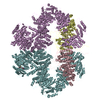
|
|---|---|
| 1 |
|
-Components
-Protein , 2 types, 4 molecules DCEF
| #1: Protein | Mass: 86533.594 Da / Num. of mol.: 2 Source method: isolated from a genetically manipulated source Source: (gene. exp.) #2: Protein | Mass: 273646.812 Da / Num. of mol.: 2 / Mutation: F2244L Source method: isolated from a genetically manipulated source Source: (gene. exp.) References: UniProt: P38111, non-specific serine/threonine protein kinase |
|---|
-Non-polymers , 4 types, 12 molecules 


| #3: Chemical |  Mass: 506.196 Da / Num. of mol.: 2 / Source method: obtained synthetically / Formula: C10H17N6O12P3 / Comment: AMP-PNP, energy-carrying molecule analogue*YM Mass: 506.196 Da / Num. of mol.: 2 / Source method: obtained synthetically / Formula: C10H17N6O12P3 / Comment: AMP-PNP, energy-carrying molecule analogue*YM#4: Chemical |  Mass: 65.409 Da / Num. of mol.: 2 / Source method: obtained synthetically / Formula: Zn Mass: 65.409 Da / Num. of mol.: 2 / Source method: obtained synthetically / Formula: Zn#5: Chemical | ChemComp-MG /  Mass: 24.305 Da / Num. of mol.: 4 / Source method: obtained synthetically / Formula: Mg Mass: 24.305 Da / Num. of mol.: 4 / Source method: obtained synthetically / Formula: Mg#6: Water | ChemComp-HOH / | Mass: 18.015 Da / Num. of mol.: 4 / Source method: isolated from a natural source / Formula: H2O |
|---|
-Details
| Has ligand of interest | N |
|---|
-Experimental details
-Experiment
| Experiment | Method: ELECTRON MICROSCOPY |
|---|---|
| EM experiment | Aggregation state: PARTICLE / 3D reconstruction method: single particle reconstruction |
- Sample preparation
| Component | Name: Mec1-Ddc2 / Type: COMPLEX Details: Mec1-Ddc2 expressed and purified from Yeast and incubated with Mg and AMP-PNP Entity ID: #1-#2 / Source: RECOMBINANT |
|---|---|
| Molecular weight | Value: 0.72 MDa / Experimental value: NO |
| Source (natural) | Organism: |
| Source (recombinant) | Organism: |
| Buffer solution | pH: 7.4 |
| Specimen | Embedding applied: NO / Shadowing applied: NO / Staining applied: NO / Vitrification applied: YES |
| Specimen support | Grid material: GOLD / Grid mesh size: 300 divisions/in. / Grid type: UltrAuFoil |
| Vitrification | Instrument: FEI VITROBOT MARK IV / Cryogen name: ETHANE / Humidity: 95 % |
- Electron microscopy imaging
| Experimental equipment |  Model: Titan Krios / Image courtesy: FEI Company |
|---|---|
| Microscopy | Model: TFS KRIOS |
| Electron gun | Electron source:  FIELD EMISSION GUN / Accelerating voltage: 300 kV / Illumination mode: FLOOD BEAM FIELD EMISSION GUN / Accelerating voltage: 300 kV / Illumination mode: FLOOD BEAM |
| Electron lens | Mode: BRIGHT FIELD / Nominal magnification: 81000 X / Cs: 2.7 mm / C2 aperture diameter: 70 µm |
| Specimen holder | Cryogen: NITROGEN / Specimen holder model: FEI TITAN KRIOS AUTOGRID HOLDER |
| Image recording | Average exposure time: 2.5 sec. / Electron dose: 51 e/Å2 / Film or detector model: GATAN K3 (6k x 4k) / Num. of grids imaged: 1 / Num. of real images: 15902 |
| Image scans | Width: 5760 / Height: 4092 |
- Processing
| Software |
| ||||||||||||||||||||||||||||||||||||||||||||
|---|---|---|---|---|---|---|---|---|---|---|---|---|---|---|---|---|---|---|---|---|---|---|---|---|---|---|---|---|---|---|---|---|---|---|---|---|---|---|---|---|---|---|---|---|---|
| EM software |
| ||||||||||||||||||||||||||||||||||||||||||||
| CTF correction | Type: PHASE FLIPPING AND AMPLITUDE CORRECTION | ||||||||||||||||||||||||||||||||||||||||||||
| Particle selection | Num. of particles selected: 900000 / Details: Template-based picking | ||||||||||||||||||||||||||||||||||||||||||||
| Symmetry | Point symmetry: C2 (2 fold cyclic) | ||||||||||||||||||||||||||||||||||||||||||||
| 3D reconstruction | Resolution: 2.82 Å / Resolution method: FSC 0.143 CUT-OFF / Num. of particles: 53581 / Num. of class averages: 1 / Symmetry type: POINT | ||||||||||||||||||||||||||||||||||||||||||||
| Atomic model building | Protocol: FLEXIBLE FIT / Space: REAL | ||||||||||||||||||||||||||||||||||||||||||||
| Atomic model building | PDB-ID: 5X6O Accession code: 5X6O / Source name: PDB / Type: experimental model | ||||||||||||||||||||||||||||||||||||||||||||
| Refinement | Stereochemistry target values: GeoStd + Monomer Library | ||||||||||||||||||||||||||||||||||||||||||||
| Refine LS restraints |
|

