 Movie
Movie Controller

[English] 日本語
 Yorodumi
- PDB-6kg3: Crystal structure of Nicotinic acid mononucleotide adenylyltransf... -
+ Open data

- Basic information
| Entry | Database: PDB / ID: 6kg3 | ||||||
|---|---|---|---|---|---|---|---|
| Title | Crystal structure of Nicotinic acid mononucleotide adenylyltransferase mutant P22K/Y84V/Y118D/C132Q/W176F from Escherichia coli | ||||||
 Components Components | Probable nicotinate-nucleotide adenylyltransferase | ||||||
 Keywords Keywords | TRANSFERASE / Nicotinic acid mononucleotide adenylyltransferase | ||||||
| Function / homology |  Function and homology information Function and homology information'de novo' NAD+ biosynthetic process from L-aspartate / nicotinamide-nucleotide adenylyltransferase activity / nicotinate-nucleotide adenylyltransferase / nicotinate-nucleotide adenylyltransferase activity / NAD+ biosynthetic process via the salvage pathway / NAD+ biosynthetic process / ATP binding Similarity search - Function | ||||||
| Biological species |  | ||||||
| Method |  X-RAY DIFFRACTION / SYNCHROTRON / MOLECULAR REPLACEMENT / Resolution: 3.08 Å X-RAY DIFFRACTION / SYNCHROTRON / MOLECULAR REPLACEMENT / Resolution: 3.08 Å | ||||||
 Authors Authors | Xue, S. / Zhao, Z. / Wang, X. / Feng, Y. | ||||||
 Citation Citation | Journal: To Be Published Title: Crystal structure of Nicotinic acid mononucleotide adenylyltransferase mutant P22K/Y84V/Y118D/C132Q/W176F from Escherichia coli Authors: Xue, S. / Feng, Y. / Zhao, Z. / Wang, X. | ||||||
| History |
|
- Structure visualization
| Structure viewer | Molecule: MolmilJmol/JSmol |
|---|
- Downloads & links
-Download
| PDBx/mmCIF format | 6kg3.cif.gz | 311.7 KB | Display | PDBx/mmCIF format |
|---|---|---|---|---|
| PDB format | pdb6kg3.ent.gz | 207.4 KB | Display | PDB format |
| PDBx/mmJSON format | 6kg3.json.gz | Tree view | PDBx/mmJSON format | |
| Others |  Other downloads Other downloads |
-Validation report
| Arichive directory | https://data.pdbj.org/pub/pdb/validation_reports/kg/6kg3ftp://data.pdbj.org/pub/pdb/validation_reports/kg/6kg3 | HTTPS FTP |
|---|
-Related structure data
| Related structure data |  1k4kS S: Starting model for refinement |
|---|---|
| Similar structure data |


-Links
 PDBj
PDBj- Assembly
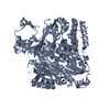
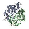
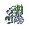
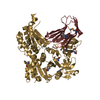
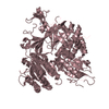
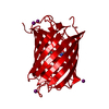
| Deposited unit | 
| ||||||||||||
|---|---|---|---|---|---|---|---|---|---|---|---|---|---|
| 1 | 
| ||||||||||||
| 2 | 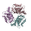
| ||||||||||||
| Unit cell |
|
-Components
| #1: Protein | Mass: 24459.758 Da / Num. of mol.: 6 / Mutation: P22K, Y84V, Y118D, C132Q, W176F Source method: isolated from a genetically manipulated source Source: (gene. exp.) References: UniProt: A0A222QGJ8, UniProt: P0A752*PLUS, nicotinate-nucleotide adenylyltransferase |
|---|
-Experimental details
-Experiment
| Experiment | Method: X-RAY DIFFRACTION / Number of used crystals: 1 |
|---|
- Sample preparation
| Crystal | Density Matthews: 3.82 Å3/Da / Density % sol: 67.76 % |
|---|---|
| Crystal grow | Temperature: 300 K / Method: vapor diffusion, hanging drop Details: 1.4M Sodium phosphate monobasic monohydrate-Potassium phosphate dibasic, pH 8.1 |
-Data collection
| Diffraction | Mean temperature: 100 K / Serial crystal experiment: N |
|---|---|
| Diffraction source | Source: SYNCHROTRON / Site: SSRF  / Beamline: BL18U1 / Wavelength: 0.9798 Å / Beamline: BL18U1 / Wavelength: 0.9798 Å |
| Detector | Type: MAR CCD 165 mm / Detector: CCD / Date: Dec 27, 2017 |
| Radiation | Protocol: SINGLE WAVELENGTH / Monochromatic (M) / Laue (L): M / Scattering type: x-ray |
| Radiation wavelength | Wavelength: 0.9798 Å / Relative weight: 1 |
| Reflection | Resolution: 3.08→50 Å / Num. obs: 37906 / % possible obs: 97.48 % / Redundancy: 4.5 % / Biso Wilson estimate: 63.51 Å2 / Rmerge(I) obs: 0.135 / Net I/σ(I): 9.05 |
| Reflection shell | Resolution: 3.08→3.15 Å / Redundancy: 4.4 % / Rmerge(I) obs: 0.557 / Mean I/σ(I) obs: 2.5 / Num. unique obs: 2771 / % possible all: 69.33 |
- Processing
| Software |
| ||||||||||||||||||||||||||||||||||||||||||||||||||||||||||||||||||||||||||||||||||||||||||||||||||
|---|---|---|---|---|---|---|---|---|---|---|---|---|---|---|---|---|---|---|---|---|---|---|---|---|---|---|---|---|---|---|---|---|---|---|---|---|---|---|---|---|---|---|---|---|---|---|---|---|---|---|---|---|---|---|---|---|---|---|---|---|---|---|---|---|---|---|---|---|---|---|---|---|---|---|---|---|---|---|---|---|---|---|---|---|---|---|---|---|---|---|---|---|---|---|---|---|---|---|---|
| Refinement | Method to determine structure: MOLECULAR REPLACEMENT Starting model: 1K4K Resolution: 3.08→45.47 Å / SU ML: 0.4592 / Cross valid method: FREE R-VALUE / σ(F): 1.35 / Phase error: 30.2449
| ||||||||||||||||||||||||||||||||||||||||||||||||||||||||||||||||||||||||||||||||||||||||||||||||||
| Solvent computation | Shrinkage radii: 0.9 Å / VDW probe radii: 1.11 Å | ||||||||||||||||||||||||||||||||||||||||||||||||||||||||||||||||||||||||||||||||||||||||||||||||||
| Displacement parameters | Biso mean: 56.05 Å2 | ||||||||||||||||||||||||||||||||||||||||||||||||||||||||||||||||||||||||||||||||||||||||||||||||||
| Refinement step | Cycle: LAST / Resolution: 3.08→45.47 Å
| ||||||||||||||||||||||||||||||||||||||||||||||||||||||||||||||||||||||||||||||||||||||||||||||||||
| Refine LS restraints |
| ||||||||||||||||||||||||||||||||||||||||||||||||||||||||||||||||||||||||||||||||||||||||||||||||||
| LS refinement shell |
|
