[English] 日本語
Yorodumi
- PDB-6k4j: Crystal Structure of the the CD9 -

+
Open data


ID or keywords:

Loading...

-
Basic information

Entry
Database: PDB / ID: 6k4j
TitleCrystal Structure of the the CD9
ComponentsCD9 antigen
KeywordsMEMBRANE PROTEIN
Function / homology
Function and homology information


Acrosome Reaction and Sperm:Oocyte Membrane Binding / myoblast fusion involved in skeletal muscle regeneration / sperm-egg recognition / fusion of sperm to egg plasma membrane involved in single fertilization / regulation of macrophage migration / paranodal junction assembly / glial cell migration / platelet alpha granule membrane / negative regulation of platelet aggregation / Uptake and function of diphtheria toxin ...Acrosome Reaction and Sperm:Oocyte Membrane Binding / myoblast fusion involved in skeletal muscle regeneration / sperm-egg recognition / fusion of sperm to egg plasma membrane involved in single fertilization / regulation of macrophage migration / paranodal junction assembly / glial cell migration / platelet alpha granule membrane / negative regulation of platelet aggregation / Uptake and function of diphtheria toxin / cellular response to low-density lipoprotein particle stimulus / clathrin-coated endocytic vesicle membrane / receptor internalization / platelet activation / endocytic vesicle membrane / extracellular vesicle / integrin binding / Platelet degranulation / cell population proliferation / cell adhesion / negative regulation of cell population proliferation / external side of plasma membrane / focal adhesion / protein-containing complex / extracellular space / extracellular exosome / membrane / plasma membrane
Similarity search - Function
CD9, extracellular domain / Tetraspanin, conserved site / Transmembrane 4 family signature. / Tetraspanin, animals / Tetraspanin, EC2 domain superfamily / Tetraspanin/Peripherin / Tetraspanin family
Similarity search - Domain/homology
NICKEL (II) ION / (2R)-2,3-dihydroxypropyl (9Z)-octadec-9-enoate / PALMITIC ACID / CD9 antigen
Similarity search - Component
Biological speciesHomo sapiens (human)
MethodX-RAY DIFFRACTION / SYNCHROTRON / SIRAS / Resolution: 2.701 Å
AuthorsUmeda, R. / Nishizawa, T. / Sato, K. / Nureki, O.
Citation
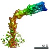
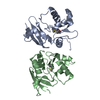
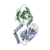
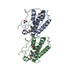
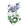
Journal: Nat Commun / Year: 2020
Title: Structural insights into tetraspanin CD9 function.
Authors: Rie Umeda / Yuhkoh Satouh / Mizuki Takemoto / Yoshiko Nakada-Nakura / Kehong Liu / Takeshi Yokoyama / Mikako Shirouzu / So Iwata / Norimichi Nomura / Ken Sato / Masahito Ikawa / Tomohiro ...Authors: Rie Umeda / Yuhkoh Satouh / Mizuki Takemoto / Yoshiko Nakada-Nakura / Kehong Liu / Takeshi Yokoyama / Mikako Shirouzu / So Iwata / Norimichi Nomura / Ken Sato / Masahito Ikawa / Tomohiro Nishizawa / Osamu Nureki /
Abstract: Tetraspanins play critical roles in various physiological processes, ranging from cell adhesion to virus infection. The members of the tetraspanin family have four membrane-spanning domains and short ...Tetraspanins play critical roles in various physiological processes, ranging from cell adhesion to virus infection. The members of the tetraspanin family have four membrane-spanning domains and short and large extracellular loops, and associate with a broad range of other functional proteins to exert cellular functions. Here we report the crystal structure of CD9 and the cryo-electron microscopic structure of CD9 in complex with its single membrane-spanning partner protein, EWI-2. The reversed cone-like molecular shape of CD9 generates membrane curvature in the crystalline lipid layers, which explains the CD9 localization in regions with high membrane curvature and its implications in membrane remodeling. The molecular interaction between CD9 and EWI-2 is mainly mediated through the small residues in the transmembrane region and protein/lipid interactions, whereas the fertilization assay revealed the critical involvement of the LEL region in the sperm-egg fusion, indicating the different dependency of each binding domain for other partner proteins.
History
DepositionMay 24, 2019Deposition site: PDBJ / Processing site: PDBJ
Revision 1.0May 13, 2020Provider: repository / Type: Initial release

-
Structure visualization

Structure viewerMolecule:
MolmilJmol/JSmol

Downloads & links

-
Assembly

Deposited unit
A: CD9 antigen
hetero molecules


Theoretical massNumber of molelcules
Total (without water)26,1124
Polymers25,4401
Non-polymers6723
Water23413
1


  • Idetical with deposited unit
  • defined by author&software
TypeNameSymmetry operationNumber
identity operation1_555x,y,z1
Buried area400 Å2
ΔGint-7 kcal/mol
Surface area13330 Å2
MethodPISA
Unit cell
Length a, b, c (Å)45.180, 125.210, 129.400
Angle α, β, γ (deg.)90.000, 90.000, 90.000
Int Tables number20
Space group name H-MC2221

-
Components

#1: Protein CD9 antigen / 5H9 antigen / Cell growth-inhibiting gene 2 protein / Leukocyte antigen MIC3 / Motility-related ...5H9 antigen / Cell growth-inhibiting gene 2 protein / Leukocyte antigen MIC3 / Motility-related protein / MRP-1 / Tetraspanin-29 / Tspan-29 / p24


Mass: 25439.947 Da / Num. of mol.: 1
Source method: isolated from a genetically manipulated source
Source: (gene. exp.) Homo sapiens (human) / Gene: CD9, MIC3, TSPAN29, GIG2 / Cell line (production host): Sf9 / Production host: 
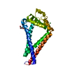
Spodoptera frugiperda (fall armyworm) / References: UniProt: P21926
#2: Chemical ChemComp-PLM / PALMITIC ACID


Mass: 256.424 Da / Num. of mol.: 1 / Source method: obtained synthetically / Formula: C16H32O2
#3: Chemical ChemComp-OLC / (2R)-2,3-dihydroxypropyl (9Z)-octadec-9-enoate / 1-Oleoyl-R-glycerol


Mass: 356.540 Da / Num. of mol.: 1 / Source method: obtained synthetically / Formula: C21H40O4
#4: Chemical ChemComp-NI / NICKEL (II) ION


Mass: 58.693 Da / Num. of mol.: 1 / Source method: obtained synthetically / Formula: Ni
#5: Water ChemComp-HOH / water


Mass: 18.015 Da / Num. of mol.: 13 / Source method: isolated from a natural source / Formula: H2O
Has ligand of interestN

-
Experimental details

-
Experiment

ExperimentMethod: X-RAY DIFFRACTION / Number of used crystals: 1

-
Sample preparation

CrystalDensity Matthews: 3.73 Å3/Da / Density % sol: 67.06 %
Crystal growTemperature: 298 K / Method: lipidic cubic phase / pH: 6.5 / Details: PEG 200

-
Data collection

DiffractionMean temperature: 100 K / Serial crystal experiment: N
Diffraction sourceSource: SYNCHROTRON / Site: SPring-8 / Beamline: BL32XU / Wavelength: 1 Å
DetectorType: DECTRIS EIGER X 9M / Detector: PIXEL / Date: Sep 21, 2016
RadiationProtocol: SINGLE WAVELENGTH / Monochromatic (M) / Laue (L): M / Scattering type: x-ray
Radiation wavelengthWavelength: 1 Å / Relative weight: 1
ReflectionResolution: 2.7→42.498 Å / Num. obs: 10431 / % possible obs: 100 % / Redundancy: 57.932 % / Biso Wilson estimate: 54.66 Å2 / CC1/2: 0.998 / Rmerge(I) obs: 0.0513 / Rrim(I) all: 0.518 / Χ2: 1.184 / Net I/σ(I): 15.83 / Num. measured all: 604284 / Scaling rejects: 364
Reflection shell

Diffraction-ID: 1

Resolution (Å)Redundancy (%)Rmerge(I) obsMean I/σ(I) obsNum. measured obsNum. possibleNum. unique obsCC1/2Rrim(I) all% possible all
2.7-2.8659.7923.1351.797042162316230.5133.162100
2.86-3.0658.9722.5212.3392409156715670.7542.543100
3.06-3.3157.6511.6323.6886650150315030.8951.646100
3.31-3.6257.0680.927.2374245130113010.9650.928100
3.62-4.0557.6680.41716.6671508124012400.9910.421100
4.05-4.6759.7890.22330.9864572108010800.9980.225100
4.67-5.7256.9690.18634.27535519409400.9980.188100
5.72-8.0853.1560.15338.96392827397390.9990.154100
8.08-42.49857.1350.09765.52250254424380.9990.09899.1

-
Processing

Software
NameVersionClassification
XSCALEdata scaling
PHENIX1.13_2998refinement
PDB_EXTRACT3.25data extraction
XDSdata reduction
PHASERphasing
RefinementMethod to determine structure: SIRAS / Resolution: 2.701→42.498 Å / SU ML: 0.48 / Cross valid method: THROUGHOUT / σ(F): 1.38 / Phase error: 30.3
RfactorNum. reflection% reflection
Rfree0.2889 1043 10 %
Rwork0.262 --
obs0.2647 10429 99.62 %
Solvent computationShrinkage radii: 0.9 Å / VDW probe radii: 1.11 Å
Displacement parametersBiso max: 124.94 Å2 / Biso mean: 58.6986 Å2 / Biso min: 30.23 Å2
Refinement stepCycle: final / Resolution: 2.701→42.498 Å
ProteinNucleic acidLigandSolventTotal
Num. atoms1673 0 30 13 1716
Biso mean--68.22 49.78 -
Num. residues----220
LS refinement shell

Refine-ID: X-RAY DIFFRACTION / Rfactor Rfree error: 0

Resolution (Å)Rfactor RfreeNum. reflection RfreeRfactor RworkNum. reflection Rwork% reflection obs (%)
2.7005-2.84290.42281420.337127897
2.8429-3.0210.34021470.29241329100
3.021-3.25410.34541470.29441316100
3.2541-3.58150.33051470.26971325100
3.5815-4.09930.26621510.24761351100
4.0993-5.16330.25611500.24511363100
5.1633-42.4980.25711590.24991424100

+
About Yorodumi

-
News

-
Feb 9, 2022. New format data for meta-information of EMDB entries

New format data for meta-information of EMDB entries

  • Version 3 of the EMDB header file is now the official format.
  • The previous official version 1.9 will be removed from the archive.

Related info.:EMDB header

External links:wwPDB to switch to version 3 of the EMDB data model

-
Aug 12, 2020. Covid-19 info

Covid-19 info

URL: https://pdbjlvh1.pdbj.org/emnavi/covid19.php

New page: Covid-19 featured information page in EM Navigator.

Related info.:Covid-19 info / Mar 5, 2020. Novel coronavirus structure data

+
Mar 5, 2020. Novel coronavirus structure data

Novel coronavirus structure data

Related info.:Yorodumi Speices / Aug 12, 2020. Covid-19 info

External links:COVID-19 featured content - PDBj / Molecule of the Month (242):Coronavirus Proteases

+
Jan 31, 2019. EMDB accession codes are about to change! (news from PDBe EMDB page)

EMDB accession codes are about to change! (news from PDBe EMDB page)

  • The allocation of 4 digits for EMDB accession codes will soon come to an end. Whilst these codes will remain in use, new EMDB accession codes will include an additional digit and will expand incrementally as the available range of codes is exhausted. The current 4-digit format prefixed with “EMD-” (i.e. EMD-XXXX) will advance to a 5-digit format (i.e. EMD-XXXXX), and so on. It is currently estimated that the 4-digit codes will be depleted around Spring 2019, at which point the 5-digit format will come into force.
  • The EM Navigator/Yorodumi systems omit the EMD- prefix.

Related info.:Q: What is EMD? / ID/Accession-code notation in Yorodumi/EM Navigator

External links:EMDB Accession Codes are Changing Soon! / Contact to PDBj

+
Jul 12, 2017. Major update of PDB

Major update of PDB

  • wwPDB released updated PDB data conforming to the new PDBx/mmCIF dictionary.
  • This is a major update changing the version number from 4 to 5, and with Remediation, in which all the entries are updated.
  • In this update, many items about electron microscopy experimental information are reorganized (e.g. em_software).
  • Now, EM Navigator and Yorodumi are based on the updated data.

External links:wwPDB Remediation / Enriched Model Files Conforming to OneDep Data Standards Now Available in the PDB FTP Archive

-
Yorodumi

Thousand views of thousand structures

  • Yorodumi is a browser for structure data from EMDB, PDB, SASBDB, etc.
  • This page is also the successor to EM Navigator detail page, and also detail information page/front-end page for Omokage search.
  • The word "yorodu" (or yorozu) is an old Japanese word meaning "ten thousand". "mi" (miru) is to see.

Related info.:EMDB / PDB / SASBDB / Comparison of 3 databanks / Yorodumi Search / Aug 31, 2016. New EM Navigator & Yorodumi / Yorodumi Papers / Jmol/JSmol / Function and homology information / Changes in new EM Navigator and Yorodumi

Read more