 Movie
Movie Controller

[English] 日本語
 Yorodumi
- PDB-4zyg: Crystal structure of methylated Sulfolobus solfataricus O6-methyl... -
+ Open data

- Basic information
| Entry | Database: PDB / ID: 4zyg | ||||||
|---|---|---|---|---|---|---|---|
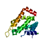
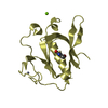
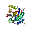
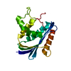
| Title | Crystal structure of methylated Sulfolobus solfataricus O6-methylguanine methyltransferase | ||||||
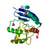
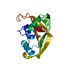
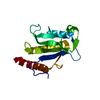
 Components Components | Methylated-DNA--protein-cysteine methyltransferase | ||||||
 Keywords Keywords | TRANSFERASE / extremophiles / DNA repair / alkylated DNA-protein alkyltransferase / methylated protein | ||||||
| Function / homology |  Function and homology information Function and homology informationmethylated-DNA-[protein]-cysteine S-methyltransferase / methylated-DNA-[protein]-cysteine S-methyltransferase activity / DNA alkylation repair / methylation / cytoplasm Similarity search - Function | ||||||
| Biological species |   Sulfolobus solfataricus (archaea) Sulfolobus solfataricus (archaea) | ||||||
| Method |  X-RAY DIFFRACTION / SYNCHROTRON / MOLECULAR REPLACEMENT / Resolution: 2.8 Å X-RAY DIFFRACTION / SYNCHROTRON / MOLECULAR REPLACEMENT / Resolution: 2.8 Å | ||||||
 Authors Authors | Miggiano, R. / Rossi, F. / Rizzi, M. | ||||||
 Citation Citation | Journal: Nucleic Acids Res. / Year: 2015 Title: Structure-function relationships governing activity and stability of a DNA alkylation damage repair thermostable protein. Authors: Perugino, G. / Miggiano, R. / Serpe, M. / Vettone, A. / Valenti, A. / Lahiri, S. / Rossi, F. / Rossi, M. / Rizzi, M. / Ciaramella, M. | ||||||
| History |
|
- Structure visualization
| Structure viewer | Molecule: MolmilJmol/JSmol |
|---|
- Downloads & links
-Download
| PDBx/mmCIF format | 4zyg.cif.gz | 70.6 KB | Display | PDBx/mmCIF format |
|---|---|---|---|---|
| PDB format | pdb4zyg.ent.gz | 52.6 KB | Display | PDB format |
| PDBx/mmJSON format | 4zyg.json.gz | Tree view | PDBx/mmJSON format | |
| Others |  Other downloads Other downloads |
-Validation report
| Arichive directory | https://data.pdbj.org/pub/pdb/validation_reports/zy/4zygftp://data.pdbj.org/pub/pdb/validation_reports/zy/4zyg | HTTPS FTP |
|---|
-Related structure data
| Related structure data |  4zydC  4zyeC  4zyhC  1wrjS S: Starting model for refinement C: citing same article ( |
|---|---|
| Similar structure data |


-Links
 PDBj
PDBj- Assembly
| Deposited unit | 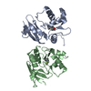
| ||||||||
|---|---|---|---|---|---|---|---|---|---|
| 1 | 
| ||||||||
| 2 | 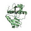
| ||||||||
| Unit cell |
|
-Components
| #1: Protein | Mass: 17074.088 Da / Num. of mol.: 2 Source method: isolated from a genetically manipulated source Source: (gene. exp.) Sulfolobus solfataricus (archaea) / Strain: ATCC 35092 / DSM 1617 / JCM 11322 / P2 / Gene: ogt, SSO2487 / Plasmid: pQE31 / Production host:  References: UniProt: Q97VW7, methylated-DNA-[protein]-cysteine S-methyltransferase #2: Water | ChemComp-HOH / |  Mass: 18.015 Da / Num. of mol.: 12 / Source method: isolated from a natural source / Formula: H2O Mass: 18.015 Da / Num. of mol.: 12 / Source method: isolated from a natural source / Formula: H2OHas protein modification | Y | |
|---|
-Experimental details
-Experiment
| Experiment | Method: X-RAY DIFFRACTION |
|---|
- Sample preparation
| Crystal | Density Matthews: 2.28 Å3/Da / Density % sol: 45.98 % |
|---|---|
| Crystal grow | Temperature: 277.15 K / Method: vapor diffusion, sitting drop Details: Ammonium acetate 0.1 M, Bis-Tris 0.1 M pH 5.5, 17% PEG 17000 PH range: 5.5 |
-Data collection
| Diffraction | Mean temperature: 100 K |
|---|---|
| Diffraction source | Source: SYNCHROTRON / Site: ESRF  / Beamline: BM30A / Wavelength: 0.972 Å / Beamline: BM30A / Wavelength: 0.972 Å |
| Detector | Type: ADSC QUANTUM 315r / Detector: CCD / Date: Feb 14, 2014 |
| Radiation | Protocol: SINGLE WAVELENGTH / Monochromatic (M) / Laue (L): M / Scattering type: x-ray |
| Radiation wavelength | Wavelength: 0.972 Å / Relative weight: 1 |
| Reflection | Resolution: 2.8→47.35 Å / Num. obs: 8764 / % possible obs: 97.5 % / Redundancy: 4 % / Rmerge(I) obs: 0.129 / Net I/σ(I): 10.1 |
| Reflection shell | Resolution: 2.8→2.95 Å / Redundancy: 4.1 % / Rmerge(I) obs: 0.481 / Mean I/σ(I) obs: 3.1 / % possible all: 98.7 |
- Processing
| Software |
| ||||||||||||||||||||||||||||
|---|---|---|---|---|---|---|---|---|---|---|---|---|---|---|---|---|---|---|---|---|---|---|---|---|---|---|---|---|---|
| Refinement | Method to determine structure: MOLECULAR REPLACEMENT Starting model: 1WRJ Resolution: 2.8→47.35 Å / SU ML: 0.4 / Cross valid method: FREE R-VALUE / σ(F): 1.35 / Phase error: 27.1 / Stereochemistry target values: ML
| ||||||||||||||||||||||||||||
| Solvent computation | Shrinkage radii: 0.9 Å / VDW probe radii: 1.11 Å / Solvent model: FLAT BULK SOLVENT MODEL | ||||||||||||||||||||||||||||
| Refinement step | Cycle: LAST / Resolution: 2.8→47.35 Å
| ||||||||||||||||||||||||||||
| Refine LS restraints |
| ||||||||||||||||||||||||||||
| LS refinement shell |
|
