 Movie
Movie Controller

[English] 日本語
 Yorodumi
- PDB-2c02: Crystal Structures of Eosinophil-derived Neurotoxin in Complex wi... -
+ Open data

- Basic information
| Entry | Database: PDB / ID: 2c02 | ||||||
|---|---|---|---|---|---|---|---|
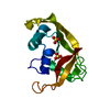
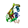
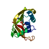
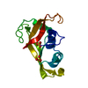
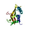
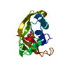
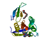
| Title | Crystal Structures of Eosinophil-derived Neurotoxin in Complex with the Inhibitors 5'-ATP, Ap3A, Ap4A and Ap5A | ||||||
 Components Components | NONSECRETORY RIBONUCLEASE | ||||||
 Keywords Keywords | HYDROLASE / ENDONUCLEASE / EOSINOPHIL / NUCLEASE / RIBONUCLEASE / RNASE US / RNASE-2 / INHIBITOR / 5'-ATP / AP3A / AP4A / AP5A / CHEMOTAXIS / GLYCOPROTEIN / POLYMORPHISM / SENSORY TRANSDUCTION | ||||||
| Function / homology |  Function and homology information Function and homology informationRNA catabolic process / pancreatic ribonuclease / ribonuclease A activity / RNA nuclease activity / innate immune response in mucosa / chemotaxis / azurophil granule lumen / defense response to virus / nucleic acid binding / hydrolase activity ...RNA catabolic process / pancreatic ribonuclease / ribonuclease A activity / RNA nuclease activity / innate immune response in mucosa / chemotaxis / azurophil granule lumen / defense response to virus / nucleic acid binding / hydrolase activity / Neutrophil degranulation / : / extracellular exosome / extracellular region Similarity search - Function | ||||||
| Biological species |  HOMO SAPIENS (human) HOMO SAPIENS (human) | ||||||
| Method |  X-RAY DIFFRACTION / MOLECULAR REPLACEMENT / Resolution: 2 Å X-RAY DIFFRACTION / MOLECULAR REPLACEMENT / Resolution: 2 Å | ||||||
 Authors Authors | Baker, M.D. / Holloway, D.E. / Swaminathan, G.J. / Acharya, K.R. | ||||||
 Citation Citation | Journal: Biochemistry / Year: 2006 Title: Crystal Structures of Eosinophil-Derived Neurotoxin (Edn) in Complex with the Inhibitors 5'- ATP, Ap(3)A, Ap(4)A, and Ap(5)A. Authors: Baker, M.D. / Holloway, D.E. / Swaminathan, G.J. / Acharya, K.R. | ||||||
| History |
| ||||||
| Remark 700 | SHEET THE SHEET STRUCTURE OF THIS MOLECULE IS BIFURCATED. IN ORDER TO REPRESENT THIS FEATURE IN ... SHEET THE SHEET STRUCTURE OF THIS MOLECULE IS BIFURCATED. IN ORDER TO REPRESENT THIS FEATURE IN THE SHEET RECORDS BELOW, TWO SHEETS ARE DEFINED. |
- Structure visualization
| Structure viewer | Molecule: MolmilJmol/JSmol |
|---|
- Downloads & links
-Download
| PDBx/mmCIF format | 2c02.cif.gz | 43.3 KB | Display | PDBx/mmCIF format |
|---|---|---|---|---|
| PDB format | pdb2c02.ent.gz | 29.4 KB | Display | PDB format |
| PDBx/mmJSON format | 2c02.json.gz | Tree view | PDBx/mmJSON format | |
| Others |  Other downloads Other downloads |
-Validation report
| Arichive directory | https://data.pdbj.org/pub/pdb/validation_reports/c0/2c02ftp://data.pdbj.org/pub/pdb/validation_reports/c0/2c02 | HTTPS FTP |
|---|
-Related structure data
| Related structure data | 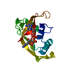 2bzzC 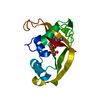 2c01C 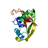 2c05C  1gqvS S: Starting model for refinement C: citing same article ( |
|---|---|
| Similar structure data |


-Links
 PDBj
PDBj


- Assembly
| Deposited unit | 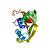
| ||||||||
|---|---|---|---|---|---|---|---|---|---|
| 1 |
| ||||||||
| Unit cell |
|
-Components
| #1: Protein | Mass: 15611.750 Da / Num. of mol.: 1 Source method: isolated from a genetically manipulated source Source: (gene. exp.) HOMO SAPIENS (human) / Cell: EOSINOPHIL / Production host:  |
|---|---|
| #2: Chemical | ChemComp-ADP /   Mass: 427.201 Da / Num. of mol.: 1 / Source method: obtained synthetically / Formula: C10H15N5O10P2 / Comment: ADP, energy-carrying molecule*YM Mass: 427.201 Da / Num. of mol.: 1 / Source method: obtained synthetically / Formula: C10H15N5O10P2 / Comment: ADP, energy-carrying molecule*YM |
| #3: Chemical | ChemComp-ACY /   Mass: 60.052 Da / Num. of mol.: 1 / Source method: obtained synthetically / Formula: C2H4O2 Mass: 60.052 Da / Num. of mol.: 1 / Source method: obtained synthetically / Formula: C2H4O2 |
| #4: Water | ChemComp-HOH /  Mass: 18.015 Da / Num. of mol.: 100 / Source method: isolated from a natural source / Formula: H2O Mass: 18.015 Da / Num. of mol.: 100 / Source method: isolated from a natural source / Formula: H2O |
| Has protein modification | Y |
-Experimental details
-Experiment
| Experiment | Method: X-RAY DIFFRACTION / Number of used crystals: 1 |
|---|
- Sample preparation
| Crystal | Density Matthews: 1.81 Å3/Da / Density % sol: 31.68 % |
|---|---|
| Crystal grow | pH: 6.5 / Details: pH 6.50 |
-Data collection
| Diffraction | Mean temperature: 203 K |
|---|---|
| Diffraction source | Source: ROTATING ANODE / Wavelength: 1.54 |
| Radiation | Protocol: SINGLE WAVELENGTH / Monochromatic (M) / Laue (L): M / Scattering type: x-ray |
| Radiation wavelength | Wavelength: 1.54 Å / Relative weight: 1 |
| Reflection | Resolution: 2→20 Å / Num. obs: 8785 / % possible obs: 99.2 % / Observed criterion σ(I): 2 / Redundancy: 3.37 % / Rmerge(I) obs: 0.05 / Net I/σ(I): 12.2 |
| Reflection shell | Resolution: 2→2.07 Å / Rmerge(I) obs: 0.32 / Mean I/σ(I) obs: 3.1 / % possible all: 99.1 |
- Processing
| Software |
| ||||||||||||||||||||||||||||||||||||||||||||||||||||||||||||
|---|---|---|---|---|---|---|---|---|---|---|---|---|---|---|---|---|---|---|---|---|---|---|---|---|---|---|---|---|---|---|---|---|---|---|---|---|---|---|---|---|---|---|---|---|---|---|---|---|---|---|---|---|---|---|---|---|---|---|---|---|---|
| Refinement | Method to determine structure: MOLECULAR REPLACEMENT Starting model: PDB ENTRY 1GQV Resolution: 2→20 Å / Cross valid method: THROUGHOUT / σ(F): 2 Details: GAMMA PHOSPHATE AND B ADENOSINE NOT MODELLED DUE TO POOR DENSITY.
| ||||||||||||||||||||||||||||||||||||||||||||||||||||||||||||
| Refinement step | Cycle: LAST / Resolution: 2→20 Å
| ||||||||||||||||||||||||||||||||||||||||||||||||||||||||||||
| Refine LS restraints |
|
