Resolution: 1.7→27.28 Å / Num. obs: 19674 / % possible obs: 98.3 % / Biso Wilson estimate: 25.34 Å2 / Rmerge(I) obs: 0.067 / Net I/σ(I): 18.38
Reflection shell
Diffraction-ID: 1,2
Resolution (Å)
Highest resolution (Å)
Rmerge(I) obs
Mean I/σ(I) obs
Num. measured obs
Num. unique obs
% possible all
1.7-1.76
0.498
2.8
13288
3373
94.7
1.76-1.83
0.425
4
18677
3564
98.9
1.83-1.91
0.366
5.3
20708
3363
95.8
1.91-2.02
0.279
7.1
25662
3773
96.9
2.02-2.14
0.185
10.7
21365
3402
99
2.14-2.31
0.133
13.7
23494
3643
98.9
2.31-2.54
0.082
21.8
25289
3589
99.9
2.54-2.9
0.07
26.2
25098
3537
99.9
2.9
0.057
37.2
34301
3636
99.9
-
Phasing
Phasing
Method: MAD
-
Processing
Software
Name
Version
Classification
NB
REFMAC
5.2.0005
refinement
XSCALE
datascaling
PDB_EXTRACT
1.701
dataextraction
XDS
datareduction
SHARP
phasing
Refinement
Method to determine structure: MAD / Resolution: 1.7→27.28 Å / Cor.coef. Fo:Fc: 0.961 / Cor.coef. Fo:Fc free: 0.939 / SU B: 5.027 / SU ML: 0.08 / TLS residual ADP flag: LIKELY RESIDUAL / Cross valid method: THROUGHOUT / σ(F): 0 / ESU R: 0.099 / ESU R Free: 0.103 Stereochemistry target values: MAXIMUM LIKELIHOOD WITH PHASES Details: 1.HYDROGENS HAVE BEEN ADDED IN THE RIDING POSITIONS. 2.ELECTRON DENSITIES FOR THE 37 N-TERMINAL RESIDUES AND FOR RESIDUES 188-198 ARE DISORDERED; THEREFORE, COORDINATES FOR ATOMS IN THESE ...Details: 1.HYDROGENS HAVE BEEN ADDED IN THE RIDING POSITIONS. 2.ELECTRON DENSITIES FOR THE 37 N-TERMINAL RESIDUES AND FOR RESIDUES 188-198 ARE DISORDERED; THEREFORE, COORDINATES FOR ATOMS IN THESE REGIONS ARE NOT INCLUDED IN THE MODEL. 3.THE DATA USED IN THE FINAL REFINEMENT WAS FROM A NATIVE CRYSTAL. THE REFINEMENT OF THE COORDINATES WAS RESTRAINED WITH THE EXPERIMENTAL PHASES FROM A CRYSTAL OF THE SELENOMETHIONINE-SUBSTITUTED PROTEIN THAT WAS USED FOR INITIAL PHASE DETERMINATION BY MULTIPLE WAVELENGTH ANOMALOUS DISPERSION.
Rfactor
Num. reflection
% reflection
Selection details
Rfree
0.22
984
5 %
RANDOM
Rwork
0.178
-
-
-
obs
0.18
19611
99.79 %
-
Solvent computation
Ion probe radii: 0.8 Å / Shrinkage radii: 0.8 Å / VDW probe radii: 1.2 Å / Solvent model: MASK
Displacement parameters
Biso mean: 19.501 Å2
Baniso -1
Baniso -2
Baniso -3
1-
-0.28 Å2
0 Å2
0 Å2
2-
-
-0.28 Å2
0 Å2
3-
-
-
0.57 Å2
Refinement step
Cycle: LAST / Resolution: 1.7→27.28 Å
Protein
Nucleic acid
Ligand
Solvent
Total
Num. atoms
1216
0
0
132
1348
Refine LS restraints
Refine-ID
Type
Dev ideal
Dev ideal target
Number
X-RAY DIFFRACTION
r_bond_refined_d
0.011
0.022
1254
X-RAY DIFFRACTION
r_bond_other_d
0.001
0.02
1210
X-RAY DIFFRACTION
r_angle_refined_deg
1.344
1.966
1713
X-RAY DIFFRACTION
r_angle_other_deg
0.78
3
2782
X-RAY DIFFRACTION
r_dihedral_angle_1_deg
6.218
5
166
X-RAY DIFFRACTION
r_dihedral_angle_2_deg
40.646
22.653
49
X-RAY DIFFRACTION
r_dihedral_angle_3_deg
10.805
15
195
X-RAY DIFFRACTION
r_dihedral_angle_4_deg
19.185
15
12
X-RAY DIFFRACTION
r_chiral_restr
0.074
0.2
205
X-RAY DIFFRACTION
r_gen_planes_refined
0.005
0.02
1414
X-RAY DIFFRACTION
r_gen_planes_other
0.001
0.02
244
X-RAY DIFFRACTION
r_nbd_refined
0.211
0.2
252
X-RAY DIFFRACTION
r_nbd_other
0.185
0.2
1267
X-RAY DIFFRACTION
r_nbtor_refined
0.169
0.2
633
X-RAY DIFFRACTION
r_nbtor_other
0.081
0.2
849
X-RAY DIFFRACTION
r_xyhbond_nbd_refined
0.147
0.2
106
X-RAY DIFFRACTION
r_symmetry_vdw_refined
0.079
0.2
11
X-RAY DIFFRACTION
r_symmetry_vdw_other
0.246
0.2
62
X-RAY DIFFRACTION
r_symmetry_hbond_refined
0.177
0.2
10
X-RAY DIFFRACTION
r_mcbond_it
1.716
3
876
X-RAY DIFFRACTION
r_mcbond_other
0.434
3
338
X-RAY DIFFRACTION
r_mcangle_it
2.317
5
1325
X-RAY DIFFRACTION
r_scbond_it
4.089
8
455
X-RAY DIFFRACTION
r_scangle_it
5.939
11
387
LS refinement shell
Resolution: 1.699→1.743 Å / Total num. of bins used: 20
Rfactor
Num. reflection
% reflection
Rfree
0.331
77
-
Rwork
0.259
1299
-
obs
-
1376
97.87 %
Refinement TLS params.
Method: refined / Origin x: -21.6381 Å / Origin y: 13.7363 Å / Origin z: 1.7575 Å
11
12
13
21
22
23
31
32
33
T
-0.0668 Å2
-0.0047 Å2
0.0176 Å2
-
-0.0323 Å2
0.0241 Å2
-
-
-0.0554 Å2
L
1.4188 °2
-0.0867 °2
0.6714 °2
-
1.0025 °2
-0.082 °2
-
-
1.311 °2
S
-0.0107 Å °
-0.0629 Å °
-0.0972 Å °
-0.102 Å °
-0.0071 Å °
-0.0744 Å °
0.0388 Å °
0.0431 Å °
0.0178 Å °
Refinement TLS group
Selection: ALL
+
About Yorodumi
-
News
-
Feb 9, 2022. New format data for meta-information of EMDB entries
New format data for meta-information of EMDB entries
Version 3 of the EMDB header file is now the official format.
The previous official version 1.9 will be removed from the archive.
In the structure databanks used in Yorodumi, some data are registered as the other names, "COVID-19 virus" and "2019-nCoV". Here are the details of the virus and the list of structure data.
Jan 31, 2019. EMDB accession codes are about to change! (news from PDBe EMDB page)
EMDB accession codes are about to change! (news from PDBe EMDB page)
The allocation of 4 digits for EMDB accession codes will soon come to an end. Whilst these codes will remain in use, new EMDB accession codes will include an additional digit and will expand incrementally as the available range of codes is exhausted. The current 4-digit format prefixed with “EMD-” (i.e. EMD-XXXX) will advance to a 5-digit format (i.e. EMD-XXXXX), and so on. It is currently estimated that the 4-digit codes will be depleted around Spring 2019, at which point the 5-digit format will come into force.
The EM Navigator/Yorodumi systems omit the EMD- prefix.
Related info.:Q: What is EMD? / ID/Accession-code notation in Yorodumi/EM Navigator
Yorodumi is a browser for structure data from EMDB, PDB, SASBDB, etc.
This page is also the successor to EM Navigator detail page, and also detail information page/front-end page for Omokage search.
The word "yorodu" (or yorozu) is an old Japanese word meaning "ten thousand". "mi" (miru) is to see.
Related info.:EMDB / PDB / SASBDB / Comparison of 3 databanks / Yorodumi Search / Aug 31, 2016. New EM Navigator & Yorodumi / Yorodumi Papers / Jmol/JSmol / Function and homology information / Changes in new EM Navigator and Yorodumi
 Movie
Movie Controller

 Yorodumi
 Open data
 Basic information
 Components
 Keywords
 Function and homology information
 Deinococcus radiodurans (radioresistant)
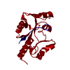
 X-RAY DIFFRACTION /
  Authors
 Citation
 Structure visualization
 Downloads & links
 Other downloads


 PDBj
PDBj Assembly
 Mass: 18.015 Da / Num. of mol.: 132 / Source method: isolated from a natural source / Formula: H2O
 Sample preparation
 Processing