Mass: 18.015 Da / Num. of mol.: 157 / Source method: isolated from a natural source / Formula: H2O
-
Experimental details
-
Experiment
Experiment
Method: X-RAY DIFFRACTION / Number of used crystals: 2
-
Sample preparation
Crystal
Density Matthews: 2.42 Å3/Da / Density % sol: 49.26 % Description: TWO DATA SETS WERE COMBINED TO OBTAIN FINAL SET. TWO CRYSTALS WERE USED FOR DATA COLLECTION. DATA FROM 15.0 -2.24A WAS COLLECTED AT CHESS A1.(CRYSTAL DIFFRACTED TO 1.7A). DATA FROM 2.24- ...Description: TWO DATA SETS WERE COMBINED TO OBTAIN FINAL SET. TWO CRYSTALS WERE USED FOR DATA COLLECTION. DATA FROM 15.0 -2.24A WAS COLLECTED AT CHESS A1.(CRYSTAL DIFFRACTED TO 1.7A). DATA FROM 2.24-1.50A WAS COLLECTED AT CHESS F1. SEE JBC (99) ARTICLE FOR DETAILS.
Crystal grow
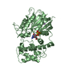
pH: 7 Details: PROTEIN BOUND TO GPPNHP WAS CRYSTALLIZED IN 1.8-2.0M (NH4)2SO3, 100MM SODIUM ACETATE, H 6.0, 1MM MGSO4, 1MM GPPNHP, AND 5MM DTT. PRIOR TO DATA COLLECTION CRYSTALS WERE TRANSFERED TO 2.0M ...Details: PROTEIN BOUND TO GPPNHP WAS CRYSTALLIZED IN 1.8-2.0M (NH4)2SO3, 100MM SODIUM ACETATE, H 6.0, 1MM MGSO4, 1MM GPPNHP, AND 5MM DTT. PRIOR TO DATA COLLECTION CRYSTALS WERE TRANSFERED TO 2.0M LI2SO4, 1MM GPPNHP, 3MM DTT, 1MM MGSO4, 100MM BES, PH 7.0, AND 15% GLYCEROL. CRYSTALS WERE FLASH FROZEN IN LIQUID PROPANE.
Crystal grow
*PLUS
pH: 8 / Method: vapor diffusion
Components of the solutions
*PLUS
ID
Conc.
Common name
Crystal-ID
Sol-ID
Chemical formula
1
10mg/ml
protein
1
drop
2
1.4M
potassiumphosphate
1
drop
3
25mM
EPPS
1
drop
4
1mM
1
drop
MgSO4
6
2.5mM
dithiothreitol
1
drop
7
10mM
EDTA
1
drop
8
100mM
1
reservoir
NaCl
9
1.45-1.65 M
potassiumphosphate
1
reservoir
5
GppNHp
1
drop
-
Data collection
Diffraction
ID
Mean temperature (K)
Crystal-ID
1
100
1
2
1
Diffraction source
Source
Site
Beamline
ID
Wavelength
SYNCHROTRON
CHESS
A1
1
0.91
SYNCHROTRON
CHESS
F1
2
0.918
Detector
Type
ID
Detector
Date
ADSC QUANTUM 4
1
CCD
Jun 1, 1998
ADSC QUANTUM 4
2
CCD
Jun 1, 1998
Radiation
ID
Protocol
Monochromatic (M) / Laue (L)
Scattering type
Wavelength-ID
1
SINGLEWAVELENGTH
M
x-ray
1
2
SINGLEWAVELENGTH
M
x-ray
2
Radiation wavelength
ID
Wavelength (Å)
Relative weight
1
0.91
1
2
0.918
1
Reflection
Resolution: 1.5→15 Å / Num. all: 57771 / Num. obs: 57771 / % possible obs: 90.2 % / Observed criterion σ(I): 0 / Redundancy: 3.92 % / Biso Wilson estimate: 15.94 Å2 / Rmerge(I) obs: 0.051 / Net I/σ(I): 21.6
Reflection shell
Resolution: 1.5→1.55 Å / Redundancy: 2.5 % / Rmerge(I) obs: 0.14 / Mean I/σ(I) obs: 7.5 / % possible all: 77.4
Reflection
*PLUS
Reflection shell
*PLUS
% possible obs: 77.4 %
-
Processing
Software
Name
Version
Classification
X-PLOR
modelbuilding
X-PLOR
3.851
refinement
DENZO
datareduction
SCALEPACK
datascaling
X-PLOR
phasing
Refinement
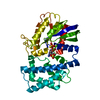
Method to determine structure: MOLECULAR REPLACEMENT Starting model: 1GIA
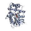
Resolution: 1.5→15 Å / Data cutoff high absF: 100000 / Data cutoff low absF: 0.1 / Isotropic thermal model: RESTRAINED / Cross valid method: THROUGHOUT / σ(F): 1 Details: RESIDUES 2 - 31, AND 348 - 354 ARE DISORDERED AND DID NOT APPEAR IN THE DENSITY. SIDE- CHAINS THAT ARE PARTIALLY DISORDERED HAVE THE OCCUPANCYS OF RELEVANT ATOMS SET TO 0.00.
Rfactor
Num. reflection
% reflection
Selection details
Rfree
0.235
2895
4.6 %
RANDOM
Rwork
0.213
-
-
-
obs
-
57291
90.84 %
-
Displacement parameters
Biso mean: 18.02 Å2
Refinement step
Cycle: LAST / Resolution: 1.5→15 Å
Protein
Nucleic acid
Ligand
Solvent
Total
Num. atoms
2543
0
33
157
2733
Refine LS restraints
Refine-ID
Type
Dev ideal
Dev ideal target
X-RAY DIFFRACTION
x_bond_d
0.011
X-RAY DIFFRACTION
x_bond_d_na
X-RAY DIFFRACTION
x_bond_d_prot
X-RAY DIFFRACTION
x_angle_d
X-RAY DIFFRACTION
x_angle_d_na
X-RAY DIFFRACTION
x_angle_d_prot
X-RAY DIFFRACTION
x_angle_deg
1.37
X-RAY DIFFRACTION
x_angle_deg_na
X-RAY DIFFRACTION
x_angle_deg_prot
X-RAY DIFFRACTION
x_dihedral_angle_d
22.83
X-RAY DIFFRACTION
x_dihedral_angle_d_na
X-RAY DIFFRACTION
x_dihedral_angle_d_prot
X-RAY DIFFRACTION
x_improper_angle_d
1.36
X-RAY DIFFRACTION
x_improper_angle_d_na
X-RAY DIFFRACTION
x_improper_angle_d_prot
X-RAY DIFFRACTION
x_mcbond_it
1.368
1.5
X-RAY DIFFRACTION
x_mcangle_it
2.166
2
X-RAY DIFFRACTION
x_scbond_it
2.619
2
X-RAY DIFFRACTION
x_scangle_it
4.17
2.5
LS refinement shell
Resolution: 1.5→1.57 Å / Total num. of bins used: 8
In the structure databanks used in Yorodumi, some data are registered as the other names, "COVID-19 virus" and "2019-nCoV". Here are the details of the virus and the list of structure data.
Jan 31, 2019. EMDB accession codes are about to change! (news from PDBe EMDB page)
EMDB accession codes are about to change! (news from PDBe EMDB page)
The allocation of 4 digits for EMDB accession codes will soon come to an end. Whilst these codes will remain in use, new EMDB accession codes will include an additional digit and will expand incrementally as the available range of codes is exhausted. The current 4-digit format prefixed with “EMD-” (i.e. EMD-XXXX) will advance to a 5-digit format (i.e. EMD-XXXXX), and so on. It is currently estimated that the 4-digit codes will be depleted around Spring 2019, at which point the 5-digit format will come into force.
The EM Navigator/Yorodumi systems omit the EMD- prefix.
Related info.:Q: What is EMD? / ID/Accession-code notation in Yorodumi/EM Navigator
Yorodumi is a browser for structure data from EMDB, PDB, SASBDB, etc.
This page is also the successor to EM Navigator detail page, and also detail information page/front-end page for Omokage search.
The word "yorodu" (or yorozu) is an old Japanese word meaning "ten thousand". "mi" (miru) is to see.
Related info.:EMDB / PDB / SASBDB / Comparison of 3 databanks / Yorodumi Search / Aug 31, 2016. New EM Navigator & Yorodumi / Yorodumi Papers / Jmol/JSmol / Function and homology information / Changes in new EM Navigator and Yorodumi
 Movie
Movie Controller

 Yorodumi
 Open data
 Basic information
 Components
 Keywords
 Function and homology information
 X-RAY DIFFRACTION /
  Authors
 Citation
 Structure visualization
 Downloads & links
 Other downloads

 PDBj
PDBj


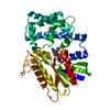
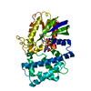
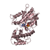
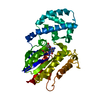
 Assembly


 Mass: 24.305 Da / Num. of mol.: 1 / Source method: obtained synthetically / Formula: Mg
 Mass: 522.196 Da / Num. of mol.: 1 / Source method: obtained synthetically / Formula: C10H17N6O13P3
 Mass: 18.015 Da / Num. of mol.: 157 / Source method: isolated from a natural source / Formula: H2O
 Sample preparation
 Processing