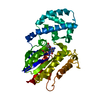
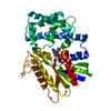
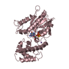
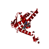
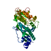
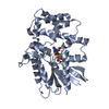
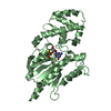
[English] 日本語
Yorodumi
- PDB-5kdl: Crystal structure of the 4 alanine insertion variant of the Gi al... -

+
Open data


ID or keywords:

Loading...

-
Basic information

Entry
Database: PDB / ID: 5kdl
TitleCrystal structure of the 4 alanine insertion variant of the Gi alpha1 subunit bound to GTPgammaS
ComponentsGuanine nucleotide-binding protein G(i) subunit alpha-1
KeywordsSIGNALING PROTEIN / Heterotrimeric G protein G protein Coupled Receptors G protein activation G protein structure GDP release
Function / homology
Function and homology information


Extra-nuclear estrogen signaling / Adenylate cyclase inhibitory pathway / Adrenaline,noradrenaline inhibits insulin secretion / ADP signalling through P2Y purinoceptor 12 / GTPase activating protein binding / negative regulation of synaptic transmission / G alpha (i) signalling events / neurotransmitter receptor localization to postsynaptic specialization membrane / positive regulation of protein localization to cell cortex / regulation of cAMP-mediated signaling ...Extra-nuclear estrogen signaling / Adenylate cyclase inhibitory pathway / Adrenaline,noradrenaline inhibits insulin secretion / ADP signalling through P2Y purinoceptor 12 / GTPase activating protein binding / negative regulation of synaptic transmission / G alpha (i) signalling events / neurotransmitter receptor localization to postsynaptic specialization membrane / positive regulation of protein localization to cell cortex / regulation of cAMP-mediated signaling / D2 dopamine receptor binding / G protein-coupled serotonin receptor binding / regulation of mitotic spindle organization / cellular response to forskolin / G protein-coupled receptor binding / G-protein beta/gamma-subunit complex binding / adenylate cyclase-modulating G protein-coupled receptor signaling pathway / GDP binding / heterotrimeric G-protein complex / cell cortex / midbody / postsynapse / G protein-coupled receptor signaling pathway / cell division / GTPase activity / centrosome / glutamatergic synapse / GTP binding / magnesium ion binding / protein-containing complex / nucleus / plasma membrane / cytoplasm
Similarity search - Function
GI Alpha 1, domain 2-like / GI Alpha 1, domain 2-like / G-protein alpha subunit, group I / G-alpha domain profile. / Guanine nucleotide binding protein (G-protein), alpha subunit / G protein alpha subunit, helical insertion / G-protein alpha subunit / G protein alpha subunit / P-loop containing nucleotide triphosphate hydrolases / Rossmann fold ...GI Alpha 1, domain 2-like / GI Alpha 1, domain 2-like / G-protein alpha subunit, group I / G-alpha domain profile. / Guanine nucleotide binding protein (G-protein), alpha subunit / G protein alpha subunit, helical insertion / G-protein alpha subunit / G protein alpha subunit / P-loop containing nucleotide triphosphate hydrolases / Rossmann fold / P-loop containing nucleoside triphosphate hydrolase / Orthogonal Bundle / 3-Layer(aba) Sandwich / Mainly Alpha / Alpha Beta
Similarity search - Domain/homology
5'-GUANOSINE-DIPHOSPHATE-MONOTHIOPHOSPHATE / Guanine nucleotide-binding protein G(i) subunit alpha-1
Similarity search - Component
Biological speciesRattus norvegicus (Norway rat)
MethodX-RAY DIFFRACTION / SYNCHROTRON / MOLECULAR REPLACEMENT / Resolution: 2.665 Å
AuthorsKaya, A.I. / Lokits, A.D. / Gilbert, J. / Iverson, T.M. / Meiler, J. / Hamm, H.E.
Funding support United States, 4items
OrganizationGrant numberCountry
National Institutes of Health/National Eye Institute (NIH/NEI)RO1 EY006062 United States
National Institutes of Health/National Institute of General Medical Sciences (NIH/NIGMS)RO1 GM120569 United States
National Institutes of Health/National Institute of General Medical Sciences (NIH/NIGMS)RO1 GM080403 United States
National Institutes of Health/National Center for Research Resources (NIH/NCRR)RO1 RR026915 United States
CitationJournal: J.Biol.Chem. / Year: 2016
Title: A Conserved Hydrophobic Core in G alpha i1 Regulates G Protein Activation and Release from Activated Receptor.
Authors: Kaya, A.I. / Lokits, A.D. / Gilbert, J.A. / Iverson, T.M. / Meiler, J. / Hamm, H.E.
History
DepositionJun 8, 2016Deposition site: RCSB / Processing site: RCSB
Revision 1.0Aug 3, 2016Provider: repository / Type: Initial release
Revision 1.1Aug 10, 2016Group: Database references
Revision 1.2Sep 21, 2016Group: Database references
Revision 1.3Sep 13, 2017Group: Author supporting evidence / Database references / Derived calculations
Category: citation / pdbx_audit_support / pdbx_struct_oper_list
Item: _citation.journal_id_CSD / _pdbx_audit_support.funding_organization / _pdbx_struct_oper_list.symmetry_operation
Revision 1.4Dec 4, 2019Group: Author supporting evidence / Category: pdbx_audit_support / Item: _pdbx_audit_support.funding_organization
Revision 1.5Sep 27, 2023Group: Data collection / Database references ...Data collection / Database references / Derived calculations / Refinement description
Category: chem_comp_atom / chem_comp_bond ...chem_comp_atom / chem_comp_bond / database_2 / pdbx_initial_refinement_model / pdbx_struct_conn_angle / struct_conn
Item: _database_2.pdbx_DOI / _database_2.pdbx_database_accession ..._database_2.pdbx_DOI / _database_2.pdbx_database_accession / _pdbx_struct_conn_angle.ptnr1_auth_comp_id / _pdbx_struct_conn_angle.ptnr1_auth_seq_id / _pdbx_struct_conn_angle.ptnr1_label_asym_id / _pdbx_struct_conn_angle.ptnr1_label_atom_id / _pdbx_struct_conn_angle.ptnr1_label_comp_id / _pdbx_struct_conn_angle.ptnr3_auth_comp_id / _pdbx_struct_conn_angle.ptnr3_auth_seq_id / _pdbx_struct_conn_angle.ptnr3_label_asym_id / _pdbx_struct_conn_angle.ptnr3_label_atom_id / _pdbx_struct_conn_angle.ptnr3_label_comp_id / _pdbx_struct_conn_angle.value / _struct_conn.pdbx_dist_value / _struct_conn.ptnr1_auth_asym_id / _struct_conn.ptnr1_auth_comp_id / _struct_conn.ptnr1_auth_seq_id / _struct_conn.ptnr1_label_asym_id / _struct_conn.ptnr1_label_atom_id / _struct_conn.ptnr1_label_comp_id / _struct_conn.ptnr1_label_seq_id / _struct_conn.ptnr2_auth_asym_id / _struct_conn.ptnr2_auth_comp_id / _struct_conn.ptnr2_auth_seq_id / _struct_conn.ptnr2_label_asym_id / _struct_conn.ptnr2_label_atom_id / _struct_conn.ptnr2_label_comp_id

-
Structure visualization

Structure viewerMolecule:
MolmilJmol/JSmol

Downloads & links

-
Assembly

Deposited unit
A: Guanine nucleotide-binding protein G(i) subunit alpha-1
B: Guanine nucleotide-binding protein G(i) subunit alpha-1
hetero molecules


Theoretical massNumber of molelcules
Total (without water)82,4946
Polymers81,3672
Non-polymers1,1274
Water724
1
A: Guanine nucleotide-binding protein G(i) subunit alpha-1
hetero molecules


Theoretical massNumber of molelcules
Total (without water)41,2473
Polymers40,6831
Non-polymers5642
Water181
TypeNameSymmetry operationNumber
identity operation1_555x,y,z1
2
B: Guanine nucleotide-binding protein G(i) subunit alpha-1
hetero molecules


Theoretical massNumber of molelcules
Total (without water)41,2473
Polymers40,6831
Non-polymers5642
Water181
TypeNameSymmetry operationNumber
identity operation1_555x,y,z1
Unit cell
Length a, b, c (Å)61.742, 77.351, 73.095
Angle α, β, γ (deg.)90.00, 99.86, 90.00
Int Tables number4
Space group name H-MP1211

-
Components

#1: Protein Guanine nucleotide-binding protein G(i) subunit alpha-1 / Adenylate cyclase-inhibiting G alpha protein


Mass: 40683.348 Da / Num. of mol.: 2
Source method: isolated from a genetically manipulated source
Source: (gene. exp.) Rattus norvegicus (Norway rat) / Gene: Gnai1, Gnai-1 / Production host: Escherichia coli (E. coli) / References: UniProt: P10824
#2: Chemical ChemComp-MG / MAGNESIUM ION


Mass: 24.305 Da / Num. of mol.: 2 / Source method: isolated from a natural source / Formula: Mg
#3: Chemical ChemComp-GSP / 5'-GUANOSINE-DIPHOSPHATE-MONOTHIOPHOSPHATE


Mass: 539.246 Da / Num. of mol.: 2 / Source method: obtained synthetically / Formula: C10H16N5O13P3S
#4: Water ChemComp-HOH / water


Mass: 18.015 Da / Num. of mol.: 4 / Source method: isolated from a natural source / Formula: H2O

-
Experimental details

-
Experiment

ExperimentMethod: X-RAY DIFFRACTION / Number of used crystals: 1

-
Sample preparation

CrystalDensity Matthews: 2.25 Å3/Da / Density % sol: 45.45 %
Crystal growTemperature: 294 K / Method: vapor diffusion, hanging drop
Details: 12-16 % polyethylene glycol (PEG) 2000 monomethyl ether, 18% 2-propanol and 100 mM MES (pH 6.0)

-
Data collection

DiffractionMean temperature: 100 K
Diffraction sourceSource: SYNCHROTRON / Site: APS / Beamline: 21-ID-F / Wavelength: 0.97 Å
DetectorType: MARMOSAIC 225 mm CCD / Detector: CCD / Date: Aug 19, 2015
RadiationProtocol: SINGLE WAVELENGTH / Monochromatic (M) / Laue (L): M / Scattering type: x-ray
Radiation wavelengthWavelength: 0.97 Å / Relative weight: 1
ReflectionResolution: 2.66→47.816 Å / Num. obs: 19135 / % possible obs: 99.5 % / Redundancy: 3.4 % / Rsym value: 0.088 / Net I/σ(I): 14.4
Reflection shellResolution: 2.66→2.8 Å / Redundancy: 3.1 % / Mean I/σ(I) obs: 2.04 / Rsym value: 0.571 / % possible all: 97.4

-
Processing

Software
NameVersionClassification
PHENIX(1.10_2155: ???)refinement
HKL-2000data reduction
HKL-2000data scaling
PHENIXphasing
RefinementMethod to determine structure: MOLECULAR REPLACEMENT
Starting model: 1GIA
Resolution: 2.665→47.816 Å / SU ML: 0.35 / Cross valid method: FREE R-VALUE / σ(F): 1.37 / Phase error: 31.54 / Stereochemistry target values: ML
RfactorNum. reflection% reflection
Rfree0.2647 1871 9.84 %
Rwork0.2093 --
obs0.2148 19008 97.39 %
Solvent computationShrinkage radii: 0.9 Å / VDW probe radii: 1.11 Å / Solvent model: FLAT BULK SOLVENT MODEL
Refinement stepCycle: LAST / Resolution: 2.665→47.816 Å
ProteinNucleic acidLigandSolventTotal
Num. atoms4960 0 66 4 5030
Refine LS restraints
Refine-IDTypeDev idealNumber
X-RAY DIFFRACTIONf_bond_d0.0035116
X-RAY DIFFRACTIONf_angle_d0.5416911
X-RAY DIFFRACTIONf_dihedral_angle_d15.8633075
X-RAY DIFFRACTIONf_chiral_restr0.038770
X-RAY DIFFRACTIONf_plane_restr0.002869
LS refinement shell
Resolution (Å)Rfactor RfreeNum. reflection RfreeRfactor RworkNum. reflection RworkRefine-ID% reflection obs (%)
2.6649-2.73690.33991010.3059934X-RAY DIFFRACTION70
2.7369-2.81750.36011360.26291363X-RAY DIFFRACTION99
2.8175-2.90840.32191570.26411320X-RAY DIFFRACTION100
2.9084-3.01230.31471480.24141331X-RAY DIFFRACTION100
3.0123-3.13290.32051510.24821345X-RAY DIFFRACTION100
3.1329-3.27550.30271300.24251348X-RAY DIFFRACTION100
3.2755-3.44810.31621520.21821359X-RAY DIFFRACTION100
3.4481-3.66410.30521500.20511358X-RAY DIFFRACTION100
3.6641-3.94680.24821250.19651353X-RAY DIFFRACTION100
3.9468-4.34380.22211410.18221353X-RAY DIFFRACTION100
4.3438-4.97180.2231490.18531359X-RAY DIFFRACTION100
4.9718-6.26170.251640.21251344X-RAY DIFFRACTION100
6.2617-47.82350.23441670.19171370X-RAY DIFFRACTION99

+
About Yorodumi

-
News

-
Feb 9, 2022. New format data for meta-information of EMDB entries

New format data for meta-information of EMDB entries

  • Version 3 of the EMDB header file is now the official format.
  • The previous official version 1.9 will be removed from the archive.

Related info.:EMDB header

External links:wwPDB to switch to version 3 of the EMDB data model

-
Aug 12, 2020. Covid-19 info

Covid-19 info

URL: https://pdbjlvh1.pdbj.org/emnavi/covid19.php

New page: Covid-19 featured information page in EM Navigator.

Related info.:Covid-19 info / Mar 5, 2020. Novel coronavirus structure data

+
Mar 5, 2020. Novel coronavirus structure data

Novel coronavirus structure data

Related info.:Yorodumi Speices / Aug 12, 2020. Covid-19 info

External links:COVID-19 featured content - PDBj / Molecule of the Month (242):Coronavirus Proteases

+
Jan 31, 2019. EMDB accession codes are about to change! (news from PDBe EMDB page)

EMDB accession codes are about to change! (news from PDBe EMDB page)

  • The allocation of 4 digits for EMDB accession codes will soon come to an end. Whilst these codes will remain in use, new EMDB accession codes will include an additional digit and will expand incrementally as the available range of codes is exhausted. The current 4-digit format prefixed with “EMD-” (i.e. EMD-XXXX) will advance to a 5-digit format (i.e. EMD-XXXXX), and so on. It is currently estimated that the 4-digit codes will be depleted around Spring 2019, at which point the 5-digit format will come into force.
  • The EM Navigator/Yorodumi systems omit the EMD- prefix.

Related info.:Q: What is EMD? / ID/Accession-code notation in Yorodumi/EM Navigator

External links:EMDB Accession Codes are Changing Soon! / Contact to PDBj

+
Jul 12, 2017. Major update of PDB

Major update of PDB

  • wwPDB released updated PDB data conforming to the new PDBx/mmCIF dictionary.
  • This is a major update changing the version number from 4 to 5, and with Remediation, in which all the entries are updated.
  • In this update, many items about electron microscopy experimental information are reorganized (e.g. em_software).
  • Now, EM Navigator and Yorodumi are based on the updated data.

External links:wwPDB Remediation / Enriched Model Files Conforming to OneDep Data Standards Now Available in the PDB FTP Archive

-
Yorodumi

Thousand views of thousand structures

  • Yorodumi is a browser for structure data from EMDB, PDB, SASBDB, etc.
  • This page is also the successor to EM Navigator detail page, and also detail information page/front-end page for Omokage search.
  • The word "yorodu" (or yorozu) is an old Japanese word meaning "ten thousand". "mi" (miru) is to see.

Related info.:EMDB / PDB / SASBDB / Comparison of 3 databanks / Yorodumi Search / Aug 31, 2016. New EM Navigator & Yorodumi / Yorodumi Papers / Jmol/JSmol / Function and homology information / Changes in new EM Navigator and Yorodumi

Read more