[English] 日本語
Yorodumi
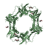
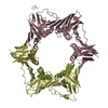
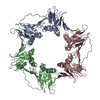
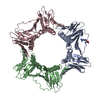
- PDB-6k2m: Crystal structure of the complex of Proliferating Cell Nuclear An... -

+
Open data


ID or keywords:

Loading...

-
Basic information

Entry
Database: PDB / ID: 6k2m
TitleCrystal structure of the complex of Proliferating Cell Nuclear Antigen from Leishmania donovani with arginine at 3.19 A resolution.
ComponentsProliferating cell nuclear antigen
KeywordsDNA BINDING PROTEIN
Function / homology
Function and homology information


PCNA complex / DNA polymerase processivity factor activity / leading strand elongation / regulation of DNA replication / mismatch repair / translesion synthesis / DNA binding
Similarity search - Function
Box / Proliferating Cell Nuclear Antigen / Proliferating Cell Nuclear Antigen - #10 / Proliferating cell nuclear antigen, PCNA, conserved site / Proliferating cell nuclear antigen signature 1. / Proliferating cell nuclear antigen, PCNA / Proliferating cell nuclear antigen, PCNA, N-terminal / Proliferating cell nuclear antigen, PCNA, C-terminal / Proliferating cell nuclear antigen, N-terminal domain / Proliferating cell nuclear antigen, C-terminal domain ...Box / Proliferating Cell Nuclear Antigen / Proliferating Cell Nuclear Antigen - #10 / Proliferating cell nuclear antigen, PCNA, conserved site / Proliferating cell nuclear antigen signature 1. / Proliferating cell nuclear antigen, PCNA / Proliferating cell nuclear antigen, PCNA, N-terminal / Proliferating cell nuclear antigen, PCNA, C-terminal / Proliferating cell nuclear antigen, N-terminal domain / Proliferating cell nuclear antigen, C-terminal domain / : / Alpha Beta
Similarity search - Domain/homology
ARGININE / Proliferating cell nuclear antigen
Similarity search - Component
Biological speciesLeishmania donovani (eukaryote)
MethodX-RAY DIFFRACTION / SYNCHROTRON / MOLECULAR REPLACEMENT / Resolution: 3.19 Å
AuthorsViswanathan, V. / Iqbal, N. / Sharma, S. / Singh, T.P.
CitationJournal: To Be Published
Title: Crystal structure of the complex of Proliferating Cell Nuclear Antigen from Leishmania donovani with arginine at 3.19 A resolution.
Authors: Viswanathan, V. / Iqbal, N. / Sharma, S. / Singh, T.P.
History
DepositionMay 15, 2019Deposition site: PDBJ / Processing site: PDBJ
Revision 1.0May 29, 2019Provider: repository / Type: Initial release
Revision 1.1Nov 22, 2023Group: Data collection / Database references / Refinement description
Category: chem_comp_atom / chem_comp_bond ...chem_comp_atom / chem_comp_bond / database_2 / pdbx_initial_refinement_model
Item: _database_2.pdbx_DOI / _database_2.pdbx_database_accession

-
Structure visualization

Structure viewerMolecule:
MolmilJmol/JSmol

Downloads & links

-
Assembly

Deposited unit
A: Proliferating cell nuclear antigen
D: Proliferating cell nuclear antigen
E: Proliferating cell nuclear antigen
B: Proliferating cell nuclear antigen
C: Proliferating cell nuclear antigen
F: Proliferating cell nuclear antigen
hetero molecules


Theoretical massNumber of molelcules
Total (without water)199,3438
Polymers198,9936
Non-polymers3502
Water1,31573
1
A: Proliferating cell nuclear antigen
B: Proliferating cell nuclear antigen
C: Proliferating cell nuclear antigen
hetero molecules


Theoretical massNumber of molelcules
Total (without water)99,6724
Polymers99,4963
Non-polymers1751
Water543
TypeNameSymmetry operationNumber
identity operation1_555x,y,z1
Buried area3750 Å2
ΔGint-14 kcal/mol
Surface area32960 Å2
MethodPISA
2
D: Proliferating cell nuclear antigen
E: Proliferating cell nuclear antigen
F: Proliferating cell nuclear antigen
hetero molecules


Theoretical massNumber of molelcules
Total (without water)99,6724
Polymers99,4963
Non-polymers1751
Water543
TypeNameSymmetry operationNumber
identity operation1_555x,y,z1
Buried area4050 Å2
ΔGint-13 kcal/mol
Surface area34140 Å2
MethodPISA
Unit cell
Length a, b, c (Å)135.926, 150.588, 171.009
Angle α, β, γ (deg.)90.00, 90.00, 90.00
Int Tables number19
Space group name H-MP212121

-
Components

#1: Protein
Proliferating cell nuclear antigen


Mass: 33165.438 Da / Num. of mol.: 6
Source method: isolated from a genetically manipulated source
Source: (gene. exp.) Leishmania donovani (eukaryote) / Gene: PCNA / Production host: Escherichia coli BL21(DE3) (bacteria) / Strain (production host): BL21(DE3) / References: UniProt: B5TV91
#2: Chemical ChemComp-ARG / ARGININE


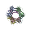
Type: L-peptide linking / Mass: 175.209 Da / Num. of mol.: 2 / Source method: obtained synthetically / Formula: C6H15N4O2 / Feature type: SUBJECT OF INVESTIGATION
#3: Water ChemComp-HOH / water


Mass: 18.015 Da / Num. of mol.: 73 / Source method: isolated from a natural source / Formula: H2O

-
Experimental details

-
Experiment

ExperimentMethod: X-RAY DIFFRACTION / Number of used crystals: 1

-
Sample preparation

CrystalDensity Matthews: 4.4 Å3/Da / Density % sol: 76.65 %
Crystal growTemperature: 298 K / Method: vapor diffusion, hanging drop / Details: 20% PEG 3350, sodium malonate

-
Data collection

DiffractionMean temperature: 100 K / Serial crystal experiment: N
Diffraction sourceSource: SYNCHROTRON / Site: ESRF / Beamline: MASSIF-1 / Wavelength: 0.966 Å
DetectorType: DECTRIS PILATUS3 2M / Detector: PIXEL / Date: Nov 8, 2018
RadiationProtocol: SINGLE WAVELENGTH / Monochromatic (M) / Laue (L): M / Scattering type: x-ray
Radiation wavelengthWavelength: 0.966 Å / Relative weight: 1
ReflectionResolution: 3.19→106.63 Å / Num. obs: 58894 / % possible obs: 99.38 % / Redundancy: 4.6 % / CC1/2: 0.999 / Rmerge(I) obs: 0.053 / Rpim(I) all: 0.028 / Rrim(I) all: 0.06 / Net I/σ(I): 16.3
Reflection shellResolution: 3.19→3.24 Å / Redundancy: 5 % / Rmerge(I) obs: 0.653 / Mean I/σ(I) obs: 2.2 / Num. unique obs: 2924 / CC1/2: 0.752 / Rpim(I) all: 0.323 / Rrim(I) all: 0.732 / % possible all: 99.8

-
Processing

Software
NameVersionClassification
REFMAC5.8.0238refinement
XDSdata reduction
XSCALEdata scaling
MOLREPphasing
Cootmodel building
RefinementMethod to determine structure: MOLECULAR REPLACEMENT
Starting model: 6J0J
Resolution: 3.19→106.63 Å / Cor.coef. Fo:Fc: 0.945 / Cor.coef. Fo:Fc free: 0.937 / SU B: 16.979 / SU ML: 0.27 / Cross valid method: THROUGHOUT / ESU R: 0.617 / ESU R Free: 0.338
RfactorNum. reflection% reflectionSelection details
Rfree0.22695 3020 5.1 %RANDOM
Rwork0.16694 ---
obs0.17011 55874 99.38 %-
Solvent computationIon probe radii: 0.8 Å / Shrinkage radii: 0.8 Å / VDW probe radii: 1.2 Å
Displacement parametersBiso mean: 110.526 Å2
Baniso -1Baniso -2Baniso -3
1-4.94 Å20 Å20 Å2
2--0.1 Å2-0 Å2
3----5.04 Å2
Refinement stepCycle: 1 / Resolution: 3.19→106.63 Å
ProteinNucleic acidLigandSolventTotal
Num. atoms11605 0 24 73 11702
Refine LS restraints
Refine-IDTypeDev idealDev ideal targetNumber
X-RAY DIFFRACTIONr_bond_refined_d0.0080.01311797
X-RAY DIFFRACTIONr_bond_other_d0.0010.01711083
X-RAY DIFFRACTIONr_angle_refined_deg1.7271.63215914
X-RAY DIFFRACTIONr_angle_other_deg1.2291.57725737
X-RAY DIFFRACTIONr_dihedral_angle_1_deg10.16951489
X-RAY DIFFRACTIONr_dihedral_angle_2_deg38.40323.247579
X-RAY DIFFRACTIONr_dihedral_angle_3_deg23.143152137
X-RAY DIFFRACTIONr_dihedral_angle_4_deg18.1661563
X-RAY DIFFRACTIONr_chiral_restr0.0690.21543
X-RAY DIFFRACTIONr_gen_planes_refined0.0070.0213130
X-RAY DIFFRACTIONr_gen_planes_other0.0020.022363
X-RAY DIFFRACTIONr_nbd_refined
X-RAY DIFFRACTIONr_nbd_other
X-RAY DIFFRACTIONr_nbtor_refined
X-RAY DIFFRACTIONr_nbtor_other
X-RAY DIFFRACTIONr_xyhbond_nbd_refined
X-RAY DIFFRACTIONr_xyhbond_nbd_other
X-RAY DIFFRACTIONr_metal_ion_refined
X-RAY DIFFRACTIONr_metal_ion_other
X-RAY DIFFRACTIONr_symmetry_vdw_refined
X-RAY DIFFRACTIONr_symmetry_vdw_other
X-RAY DIFFRACTIONr_symmetry_hbond_refined
X-RAY DIFFRACTIONr_symmetry_hbond_other
X-RAY DIFFRACTIONr_symmetry_metal_ion_refined
X-RAY DIFFRACTIONr_symmetry_metal_ion_other
X-RAY DIFFRACTIONr_mcbond_it10.27811.3745998
X-RAY DIFFRACTIONr_mcbond_other10.27611.3735997
X-RAY DIFFRACTIONr_mcangle_it14.78317.0347473
X-RAY DIFFRACTIONr_mcangle_other14.78217.0347474
X-RAY DIFFRACTIONr_scbond_it11.58612.4715799
X-RAY DIFFRACTIONr_scbond_other11.58512.475799
X-RAY DIFFRACTIONr_scangle_it
X-RAY DIFFRACTIONr_scangle_other17.33618.2498441
X-RAY DIFFRACTIONr_long_range_B_refined20.60112483
X-RAY DIFFRACTIONr_long_range_B_other20.612484
X-RAY DIFFRACTIONr_rigid_bond_restr
X-RAY DIFFRACTIONr_sphericity_free
X-RAY DIFFRACTIONr_sphericity_bonded
LS refinement shellResolution: 3.188→3.27 Å / Total num. of bins used: 20
RfactorNum. reflection% reflection
Rfree0.4 208 -
Rwork0.331 4114 -
obs--99.72 %

+
About Yorodumi

-
News

-
Feb 9, 2022. New format data for meta-information of EMDB entries

New format data for meta-information of EMDB entries

  • Version 3 of the EMDB header file is now the official format.
  • The previous official version 1.9 will be removed from the archive.

Related info.:EMDB header

External links:wwPDB to switch to version 3 of the EMDB data model

-
Aug 12, 2020. Covid-19 info

Covid-19 info

URL: https://pdbjlvh1.pdbj.org/emnavi/covid19.php

New page: Covid-19 featured information page in EM Navigator.

Related info.:Covid-19 info / Mar 5, 2020. Novel coronavirus structure data

+
Mar 5, 2020. Novel coronavirus structure data

Novel coronavirus structure data

Related info.:Yorodumi Speices / Aug 12, 2020. Covid-19 info

External links:COVID-19 featured content - PDBj / Molecule of the Month (242):Coronavirus Proteases

+
Jan 31, 2019. EMDB accession codes are about to change! (news from PDBe EMDB page)

EMDB accession codes are about to change! (news from PDBe EMDB page)

  • The allocation of 4 digits for EMDB accession codes will soon come to an end. Whilst these codes will remain in use, new EMDB accession codes will include an additional digit and will expand incrementally as the available range of codes is exhausted. The current 4-digit format prefixed with “EMD-” (i.e. EMD-XXXX) will advance to a 5-digit format (i.e. EMD-XXXXX), and so on. It is currently estimated that the 4-digit codes will be depleted around Spring 2019, at which point the 5-digit format will come into force.
  • The EM Navigator/Yorodumi systems omit the EMD- prefix.

Related info.:Q: What is EMD? / ID/Accession-code notation in Yorodumi/EM Navigator

External links:EMDB Accession Codes are Changing Soon! / Contact to PDBj

+
Jul 12, 2017. Major update of PDB

Major update of PDB

  • wwPDB released updated PDB data conforming to the new PDBx/mmCIF dictionary.
  • This is a major update changing the version number from 4 to 5, and with Remediation, in which all the entries are updated.
  • In this update, many items about electron microscopy experimental information are reorganized (e.g. em_software).
  • Now, EM Navigator and Yorodumi are based on the updated data.

External links:wwPDB Remediation / Enriched Model Files Conforming to OneDep Data Standards Now Available in the PDB FTP Archive

-
Yorodumi

Thousand views of thousand structures

  • Yorodumi is a browser for structure data from EMDB, PDB, SASBDB, etc.
  • This page is also the successor to EM Navigator detail page, and also detail information page/front-end page for Omokage search.
  • The word "yorodu" (or yorozu) is an old Japanese word meaning "ten thousand". "mi" (miru) is to see.

Related info.:EMDB / PDB / SASBDB / Comparison of 3 databanks / Yorodumi Search / Aug 31, 2016. New EM Navigator & Yorodumi / Yorodumi Papers / Jmol/JSmol / Function and homology information / Changes in new EM Navigator and Yorodumi

Read more