 Movie
Movie Controller

[English] 日本語
 Yorodumi
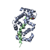
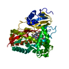
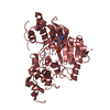
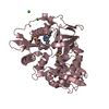
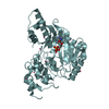
- PDB-6jwf: Holo form of Pyranose Dehydrogenase PQQ domain from Coprinopsis c... -
+ Open data

- Basic information
| Entry | Database: PDB / ID: 6jwf | ||||||
|---|---|---|---|---|---|---|---|
| Title | Holo form of Pyranose Dehydrogenase PQQ domain from Coprinopsis cinerea | ||||||
 Components Components | Extracellular PQQ-dependent sugar dehydrogenase | ||||||
 Keywords Keywords | OXIDOREDUCTASE / pyrroloquinoline quinone / sugar dehydrogenase / AA family 12 | ||||||
| Function / homology |  Function and homology information Function and homology informationOxidoreductases; Acting on the CH-OH group of donors; With unknown physiological acceptors / cellulose binding / carbohydrate metabolic process / oxidoreductase activity / extracellular region / metal ion binding Similarity search - Function | ||||||
| Biological species |  Coprinopsis cinerea (fungus) Coprinopsis cinerea (fungus) | ||||||
| Method |  X-RAY DIFFRACTION / SYNCHROTRON / MOLECULAR REPLACEMENT / Resolution: 1.3 Å X-RAY DIFFRACTION / SYNCHROTRON / MOLECULAR REPLACEMENT / Resolution: 1.3 Å | ||||||
 Authors Authors | Takeda, K. / Ishida, T. / Yoshida, M. / Samejima, M. / Ohno, H. / Igarashi, K. / Nakamura, N. | ||||||
| Funding support |  Japan, 1items Japan, 1items
| ||||||
 Citation Citation | Journal: Appl.Environ.Microbiol. / Year: 2019 Title: Crystal Structure of the Catalytic and CytochromebDomains in a Eukaryotic Pyrroloquinoline Quinone-Dependent Dehydrogenase. Authors: Takeda, K. / Ishida, T. / Yoshida, M. / Samejima, M. / Ohno, H. / Igarashi, K. / Nakamura, N. #1: Journal: To Be PublishedTitle: The first crystal structure of the catalytic domain and the cytochrome b domain in a eukaryotic PQQ-dependent dehydrogenase Authors: Takeda, K. / Ishida, T. / Yoshida, M. / Samejima, M. / Ohno, H. / Igarashi, K. / Nakamura, N. | ||||||
| History |
|
- Structure visualization
| Structure viewer | Molecule: MolmilJmol/JSmol |
|---|
- Downloads & links
-Download
| PDBx/mmCIF format | 6jwf.cif.gz | 221.2 KB | Display | PDBx/mmCIF format |
|---|---|---|---|---|
| PDB format | pdb6jwf.ent.gz | 173.1 KB | Display | PDB format |
| PDBx/mmJSON format | 6jwf.json.gz | Tree view | PDBx/mmJSON format | |
| Others |  Other downloads Other downloads |
-Validation report
| Arichive directory | https://data.pdbj.org/pub/pdb/validation_reports/jw/6jwfftp://data.pdbj.org/pub/pdb/validation_reports/jw/6jwf | HTTPS FTP |
|---|
-Related structure data
| Related structure data |  6jt5SC 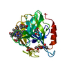 6jt6C S: Starting model for refinement C: citing same article ( |
|---|---|
| Similar structure data |


-Links
 PDBj
PDBj


- Assembly
| Deposited unit | 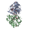
| ||||||||
|---|---|---|---|---|---|---|---|---|---|
| 1 | 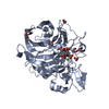
| ||||||||
| 2 | 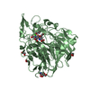
| ||||||||
| Unit cell |
|
-Components
-Protein / Sugars , 2 types, 3 molecules AB
| #1: Protein | Mass: 45173.996 Da / Num. of mol.: 2 / Fragment: PQQ dependent catalytic domain Source method: isolated from a genetically manipulated source Source: (gene. exp.) Coprinopsis cinerea (fungus) / Gene: CcSDH / Production host: Komagataella pastoris (fungus) / Strain (production host): KM71H / References: UniProt: A0A0A8IDB7, UniProt: A8P0V4*PLUS#6: Sugar | ChemComp-NAG / |  Type: D-saccharide, beta linking / Mass: 221.208 Da / Num. of mol.: 1 Type: D-saccharide, beta linking / Mass: 221.208 Da / Num. of mol.: 1Source method: isolated from a genetically manipulated source Formula: C8H15NO6 |
|---|
-Non-polymers , 5 types, 1537 molecules 
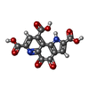


| #2: Chemical |  Mass: 40.078 Da / Num. of mol.: 2 / Source method: obtained synthetically / Formula: Ca Mass: 40.078 Da / Num. of mol.: 2 / Source method: obtained synthetically / Formula: Ca#3: Chemical |  Mass: 330.206 Da / Num. of mol.: 2 / Source method: obtained synthetically / Formula: C14H6N2O8 Mass: 330.206 Da / Num. of mol.: 2 / Source method: obtained synthetically / Formula: C14H6N2O8#4: Chemical | ChemComp-EDO /  Mass: 62.068 Da / Num. of mol.: 11 / Source method: obtained synthetically / Formula: C2H6O2 Mass: 62.068 Da / Num. of mol.: 11 / Source method: obtained synthetically / Formula: C2H6O2#5: Chemical |  Mass: 59.044 Da / Num. of mol.: 2 / Source method: obtained synthetically / Formula: C2H3O2 Mass: 59.044 Da / Num. of mol.: 2 / Source method: obtained synthetically / Formula: C2H3O2#7: Water | ChemComp-HOH / | Mass: 18.015 Da / Num. of mol.: 1520 / Source method: isolated from a natural source / Formula: H2O |
|---|
-Details
| Has protein modification | Y |
|---|
-Experimental details
-Experiment
| Experiment | Method: X-RAY DIFFRACTION / Number of used crystals: 1 |
|---|
- Sample preparation
| Crystal | Density Matthews: 2.4 Å3/Da / Density % sol: 48.71 % |
|---|---|
| Crystal grow | Temperature: 277 K / Method: batch mode / pH: 5 / Details: 5mM sodium acetate |
-Data collection
| Diffraction | Mean temperature: 100 K / Serial crystal experiment: N |
|---|---|
| Diffraction source | Source: SYNCHROTRON / Site: SPring-8 / Beamline: BL41XU / Wavelength: 0.9 Å |
| Detector | Type: DECTRIS PILATUS3 S 6M / Detector: PIXEL / Date: Jul 27, 2017 |
| Radiation | Protocol: SINGLE WAVELENGTH / Monochromatic (M) / Laue (L): M / Scattering type: x-ray |
| Radiation wavelength | Wavelength: 0.9 Å / Relative weight: 1 |
| Reflection | Resolution: 1.3→100 Å / Num. obs: 213104 / % possible obs: 99.4 % / Redundancy: 7.4 % / CC1/2: 0.999 / Rmerge(I) obs: 0.067 / Net I/σ(I): 15 |
| Reflection shell | Resolution: 1.3→1.38 Å / Redundancy: 7.2 % / Rmerge(I) obs: 0.543 / Mean I/σ(I) obs: 2.99 / Num. unique obs: 33130 / CC1/2: 0.863 / % possible all: 96.3 |
- Processing
| Software |
| ||||||||||||||||||||
|---|---|---|---|---|---|---|---|---|---|---|---|---|---|---|---|---|---|---|---|---|---|
| Refinement | Method to determine structure: MOLECULAR REPLACEMENT Starting model: 6JT5 Resolution: 1.3→71.07 Å / Cross valid method: FREE R-VALUE
| ||||||||||||||||||||
| Refinement step | Cycle: LAST / Resolution: 1.3→71.07 Å
| ||||||||||||||||||||
| LS refinement shell | Resolution: 1.297→1.33 Å / Total num. of bins used: 20
|
