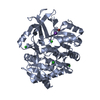
 Movie
Movie Controller


+ Open data

- Basic information
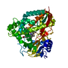
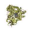
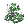
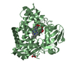
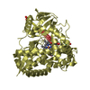
| Entry | Database: PDB / ID: 4dnj | ||||||
|---|---|---|---|---|---|---|---|
| Title | The crystal structures of 4-methoxybenzoate bound CYP199A2 | ||||||
 Components Components | Putative cytochrome P450 | ||||||
 Keywords Keywords | OXIDOREDUCTASE | ||||||
| Function / homology |  Function and homology information Function and homology information4-methoxybenzoate monooxygenase (O-demethylating) / 4-methoxybenzoate monooxygenase (O-demethylating) activity / xenobiotic metabolic process / iron ion binding / heme binding / cytoplasm Similarity search - Function | ||||||
| Biological species |  Rhodopseudomonas palustris (phototrophic) Rhodopseudomonas palustris (phototrophic) | ||||||
| Method |  X-RAY DIFFRACTION / MOLECULAR REPLACEMENT / Resolution: 1.8 Å X-RAY DIFFRACTION / MOLECULAR REPLACEMENT / Resolution: 1.8 Å | ||||||
 Authors Authors | Zhou, W. / Bell, S.G. / Yang, W. / Tan, A.B.H. / Zhou, R. / Johnson, E.O.D. / Zhang, A. / Rao, Z. / Wong, L.-L. | ||||||
 Citation Citation | Journal: Dalton Trans / Year: 2012 Title: The crystal structures of 4-methoxybenzoate bound CYP199A2 and CYP199A4: structural changes on substrate binding and the identification of an anion binding site Authors: Bell, S.G. / Yang, W. / Tan, A.B.H. / Zhou, R. / Johnson, E.O.D. / Zhang, A. / Zhou, W. / Rao, Z. / Wong, L.-L. | ||||||
| History |
|
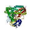
- Structure visualization
| Structure viewer | Molecule: MolmilJmol/JSmol |
|---|
- Downloads & links
-Download
| PDBx/mmCIF format | 4dnj.cif.gz | 105.8 KB | Display | PDBx/mmCIF format |
|---|---|---|---|---|
| PDB format | pdb4dnj.ent.gz | 77.8 KB | Display | PDB format |
| PDBx/mmJSON format | 4dnj.json.gz | Tree view | PDBx/mmJSON format | |
| Others |  Other downloads Other downloads |
-Validation report
| Arichive directory | https://data.pdbj.org/pub/pdb/validation_reports/dn/4dnjftp://data.pdbj.org/pub/pdb/validation_reports/dn/4dnj | HTTPS FTP |
|---|
-Related structure data
| Related structure data |  4dnzC 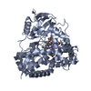 4do1C 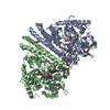 2rf7S C: citing same article ( S: Starting model for refinement |
|---|---|
| Similar structure data |


-Links
 PDBj
PDBj- Assembly
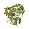
| Deposited unit | 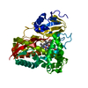
| ||||||||
|---|---|---|---|---|---|---|---|---|---|
| 1 |
| ||||||||
| Unit cell |
|
-Components
| #1: Protein | Mass: 44672.625 Da / Num. of mol.: 1 Source method: isolated from a genetically manipulated source Source: (gene. exp.) Rhodopseudomonas palustris (phototrophic)Strain: CGA009 / Gene: RPA1871 / Plasmid: pET28a / Production host: References: UniProt: Q6N8N2, Oxidoreductases; Acting on paired donors, with incorporation or reduction of molecular oxygen |
|---|---|
| #2: Chemical | ChemComp-HEM /   Mass: 616.487 Da / Num. of mol.: 1 / Source method: obtained synthetically / Formula: C34H32FeN4O4 Mass: 616.487 Da / Num. of mol.: 1 / Source method: obtained synthetically / Formula: C34H32FeN4O4 |
| #3: Chemical | ChemComp-ANN /   Mass: 152.147 Da / Num. of mol.: 1 / Source method: obtained synthetically / Formula: C8H8O3 Mass: 152.147 Da / Num. of mol.: 1 / Source method: obtained synthetically / Formula: C8H8O3 |
| #4: Chemical | ChemComp-CL /   Mass: 35.453 Da / Num. of mol.: 1 / Source method: obtained synthetically / Formula: Cl Mass: 35.453 Da / Num. of mol.: 1 / Source method: obtained synthetically / Formula: Cl |
| #5: Water | ChemComp-HOH /  Mass: 18.015 Da / Num. of mol.: 551 / Source method: isolated from a natural source / Formula: H2O Mass: 18.015 Da / Num. of mol.: 551 / Source method: isolated from a natural source / Formula: H2O |
-Experimental details
-Experiment
| Experiment | Method: X-RAY DIFFRACTION / Number of used crystals: 1 |
|---|
- Sample preparation
| Crystal | Density Matthews: 2.16 Å3/Da / Density % sol: 43.05 % |
|---|---|
| Crystal grow | Temperature: 293 K / Method: vapor diffusion, hanging drop / pH: 8.4 Details: 0.1M Tris, 26% PEG 4000, 10% isopropanol, pH 8.4, VAPOR DIFFUSION, HANGING DROP, temperature 293K |
-Data collection
| Diffraction | Mean temperature: 100 K |
|---|---|
| Diffraction source | Source: ROTATING ANODE / Type: RIGAKU MICROMAX-007 / Wavelength: 1.5418 Å |
| Detector | Type: RIGAKU RAXIS HTC / Detector: IMAGE PLATE |
| Radiation | Protocol: SINGLE WAVELENGTH / Monochromatic (M) / Laue (L): M / Scattering type: x-ray |
| Radiation wavelength | Wavelength: 1.5418 Å / Relative weight: 1 |
| Reflection | Resolution: 1.8→50 Å / Num. all: 36699 / Num. obs: 36209 / % possible obs: 98.7 % / Observed criterion σ(F): 1 / Observed criterion σ(I): 1 / Redundancy: 5 % / Biso Wilson estimate: 15.577 Å2 / Rmerge(I) obs: 0.077 / Rsym value: 0.063 / Net I/σ(I): 25.2 |
| Reflection shell | Resolution: 1.8→1.86 Å / Redundancy: 5.1 % / Rmerge(I) obs: 0.264 / Mean I/σ(I) obs: 6.64 / Num. unique all: 3521 / Rsym value: 0.248 / % possible all: 97.8 |
- Processing
| Software |
| |||||||||||||||||||||||||||||||||||||||||||||||||||||||||||||||||
|---|---|---|---|---|---|---|---|---|---|---|---|---|---|---|---|---|---|---|---|---|---|---|---|---|---|---|---|---|---|---|---|---|---|---|---|---|---|---|---|---|---|---|---|---|---|---|---|---|---|---|---|---|---|---|---|---|---|---|---|---|---|---|---|---|---|---|
| Refinement | Method to determine structure: MOLECULAR REPLACEMENT Starting model: 2RF7 Resolution: 1.8→29.07 Å / Cor.coef. Fo:Fc: 0.961 / Cor.coef. Fo:Fc free: 0.936 / SU B: 2.342 / SU ML: 0.074 / Cross valid method: THROUGHOUT / σ(F): 0 / σ(I): 0 / ESU R: 0.128 / ESU R Free: 0.125 / Stereochemistry target values: MAXIMUM LIKELIHOOD / Details: HYDROGENS HAVE BEEN ADDED IN THE RIDING POSITIONS
| |||||||||||||||||||||||||||||||||||||||||||||||||||||||||||||||||
| Solvent computation | Ion probe radii: 0.8 Å / Shrinkage radii: 0.8 Å / VDW probe radii: 1.4 Å / Solvent model: MASK | |||||||||||||||||||||||||||||||||||||||||||||||||||||||||||||||||
| Displacement parameters | Biso mean: 15.577 Å2
| |||||||||||||||||||||||||||||||||||||||||||||||||||||||||||||||||
| Refinement step | Cycle: LAST / Resolution: 1.8→29.07 Å
| |||||||||||||||||||||||||||||||||||||||||||||||||||||||||||||||||
| Refine LS restraints |
| |||||||||||||||||||||||||||||||||||||||||||||||||||||||||||||||||
| LS refinement shell | Resolution: 1.799→1.846 Å / Rfactor Rfree error: 0.125 / Total num. of bins used: 20
|
