 Movie
Movie Controller


+ Open data

- Basic information
| Entry | Database: PDB / ID: 3ofu | ||||||
|---|---|---|---|---|---|---|---|
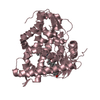
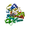
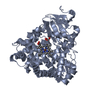
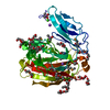
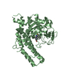
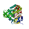
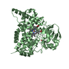
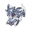
| Title | Crystal Structure of Cytochrome P450 CYP101C1 | ||||||
 Components Components | Cytochrome P450 | ||||||
 Keywords Keywords | OXIDOREDUCTASE | ||||||
| Function / homology |  Function and homology information Function and homology informationoxidoreductase activity, acting on paired donors, with incorporation or reduction of molecular oxygen / monooxygenase activity / iron ion binding / heme binding Similarity search - Function | ||||||
| Biological species |  Novosphingobium aromaticivorans (bacteria) Novosphingobium aromaticivorans (bacteria) | ||||||
| Method |  X-RAY DIFFRACTION / MOLECULAR REPLACEMENT / Resolution: 2.8 Å X-RAY DIFFRACTION / MOLECULAR REPLACEMENT / Resolution: 2.8 Å | ||||||
 Authors Authors | Zhou, W. / Ma, M. / Bell, S.G. / Yang, W. / Hao, Y. / Rees, N.H. / Bartlam, M. / Wong, L.-L. / Rao, Z. | ||||||
 Citation Citation | Journal: Chembiochem / Year: 2011 Title: Structural Analysis of CYP101C1 from Novosphingobium aromaticivorans DSM12444. Authors: Ma, M. / Bell, S.G. / Yang, W. / Hao, Y. / Rees, N.H. / Bartlam, M. / Zhou, W. / Wong, L.L. / Rao, Z. | ||||||
| History |
|
- Structure visualization
| Structure viewer | Molecule: MolmilJmol/JSmol |
|---|
- Downloads & links
-Download
| PDBx/mmCIF format | 3ofu.cif.gz | 464.4 KB | Display | PDBx/mmCIF format |
|---|---|---|---|---|
| PDB format | pdb3ofu.ent.gz | 381.4 KB | Display | PDB format |
| PDBx/mmJSON format | 3ofu.json.gz | Tree view | PDBx/mmJSON format | |
| Others |  Other downloads Other downloads |
-Validation report
| Arichive directory | https://data.pdbj.org/pub/pdb/validation_reports/of/3ofuftp://data.pdbj.org/pub/pdb/validation_reports/of/3ofu | HTTPS FTP |
|---|
-Related structure data
| Related structure data |  3oftC  2h7rS C: citing same article ( S: Starting model for refinement |
|---|---|
| Similar structure data |


-Links
 PDBj
PDBj
- Assembly
| Deposited unit | 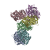
| ||||||||
|---|---|---|---|---|---|---|---|---|---|
| 1 | 
| ||||||||
| 2 | 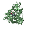
| ||||||||
| 3 | 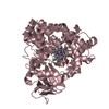
| ||||||||
| 4 | 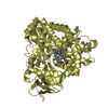
| ||||||||
| 5 | 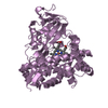
| ||||||||
| 6 | 
| ||||||||
| Unit cell |
|
-Components
| #1: Protein | Mass: 43701.445 Da / Num. of mol.: 6 Source method: isolated from a genetically manipulated source Source: (gene. exp.) Novosphingobium aromaticivorans (bacteria)Strain: DSM 12444 / Gene: Saro_2249 / Plasmid: pET28a / Production host:   Spodoptera frugiperda (fall armyworm) / Strain (production host): BL21(DE3) / References: UniProt: Q2G637 Spodoptera frugiperda (fall armyworm) / Strain (production host): BL21(DE3) / References: UniProt: Q2G637#2: Chemical | ChemComp-HEM /   Mass: 616.487 Da / Num. of mol.: 6 / Source method: obtained synthetically / Formula: C34H32FeN4O4 Mass: 616.487 Da / Num. of mol.: 6 / Source method: obtained synthetically / Formula: C34H32FeN4O4#3: Chemical |   Mass: 192.297 Da / Num. of mol.: 3 / Source method: obtained synthetically / Formula: C13H20O Mass: 192.297 Da / Num. of mol.: 3 / Source method: obtained synthetically / Formula: C13H20O#4: Water | ChemComp-HOH / |  Mass: 18.015 Da / Num. of mol.: 310 / Source method: isolated from a natural source / Formula: H2O Mass: 18.015 Da / Num. of mol.: 310 / Source method: isolated from a natural source / Formula: H2O |
|---|
-Experimental details
-Experiment
| Experiment | Method: X-RAY DIFFRACTION / Number of used crystals: 1 |
|---|
- Sample preparation
| Crystal | Density Matthews: 2.22 Å3/Da / Density % sol: 44.62 % |
|---|---|
| Crystal grow | Temperature: 289 K / Method: vapor diffusion, hanging drop / pH: 7.4 Details: 0.1M Bis-Tris, pH 5.0, 30% w/v polyethylene glycol monomethyl ether 2000, VAPOR DIFFUSION, HANGING DROP, temperature 289K |
-Data collection
| Diffraction | Mean temperature: 100 K |
|---|---|
| Diffraction source | Source: ROTATING ANODE / Type: RIGAKU MICROMAX-007 / Wavelength: 1.5418 Å |
| Detector | Type: RIGAKU RAXIS IV++ / Detector: IMAGE PLATE / Date: Sep 30, 2009 / Details: mirrors |
| Radiation | Monochromator: GRAPHITE / Protocol: SINGLE WAVELENGTH / Monochromatic (M) / Laue (L): M / Scattering type: x-ray |
| Radiation wavelength | Wavelength: 1.5418 Å / Relative weight: 1 |
| Reflection | Resolution: 2.8→150.76 Å / Num. all: 55970 / Num. obs: 53086 / % possible obs: 94.8 % / Observed criterion σ(F): 1 / Observed criterion σ(I): 1 / Redundancy: 5.3 % / Biso Wilson estimate: 27.444 Å2 / Rmerge(I) obs: 0.171 / Rsym value: 0.116 / Net I/σ(I): 13.2 |
| Reflection shell | Resolution: 2.8→2.9 Å / Redundancy: 5.3 % / Rmerge(I) obs: 0.519 / Mean I/σ(I) obs: 3.78 / Num. unique all: 5614 / Rsym value: 0.409 / % possible all: 99 |
- Processing
| Software |
| |||||||||||||||||||||||||||||||||||||||||||||||||||||||||||||||||
|---|---|---|---|---|---|---|---|---|---|---|---|---|---|---|---|---|---|---|---|---|---|---|---|---|---|---|---|---|---|---|---|---|---|---|---|---|---|---|---|---|---|---|---|---|---|---|---|---|---|---|---|---|---|---|---|---|---|---|---|---|---|---|---|---|---|---|
| Refinement | Method to determine structure: MOLECULAR REPLACEMENT Starting model: 2H7R Resolution: 2.8→50 Å / Cor.coef. Fo:Fc: 0.941 / Cor.coef. Fo:Fc free: 0.848 / SU B: 19.253 / SU ML: 0.383 / Isotropic thermal model: Overall / Cross valid method: THROUGHOUT / σ(F): 0 / σ(I): 0 / ESU R Free: 0.517 / Stereochemistry target values: MAXIMUM LIKELIHOOD / Details: HYDROGENS HAVE BEEN ADDED IN THE RIDING POSITIONS
| |||||||||||||||||||||||||||||||||||||||||||||||||||||||||||||||||
| Solvent computation | Ion probe radii: 0.8 Å / Shrinkage radii: 0.8 Å / VDW probe radii: 1.2 Å / Solvent model: MASK | |||||||||||||||||||||||||||||||||||||||||||||||||||||||||||||||||
| Displacement parameters | Biso mean: 27.444 Å2
| |||||||||||||||||||||||||||||||||||||||||||||||||||||||||||||||||
| Refine analyze | Luzzati coordinate error obs: 0.3368 Å | |||||||||||||||||||||||||||||||||||||||||||||||||||||||||||||||||
| Refinement step | Cycle: LAST / Resolution: 2.8→50 Å
| |||||||||||||||||||||||||||||||||||||||||||||||||||||||||||||||||
| Refine LS restraints |
| |||||||||||||||||||||||||||||||||||||||||||||||||||||||||||||||||
| LS refinement shell | Resolution: 2.798→2.871 Å / Rfactor Rfree error: 0.0507 / Total num. of bins used: 20
|
