Mass: 18.015 Da / Num. of mol.: 251 / Source method: isolated from a natural source / Formula: H2O
-
Details
Has protein modification
Y
Sequence details
SEQUENCE THIS CONSTRUCT (RESIDUES 21-361) WAS EXPRESSED WITH A PURIFICATION TAG MGSDKIHHHHHHENLYFQG. ...SEQUENCE THIS CONSTRUCT (RESIDUES 21-361) WAS EXPRESSED WITH A PURIFICATION TAG MGSDKIHHHHHHENLYFQG. THE TAG WAS REMOVED WITH TEV PROTEASE LEAVING ONLY A GLYCINE (0) FOLLOWED BY THE TARGET SEQUENCE.
-
Experimental details
-
Experiment
Experiment
Method: X-RAY DIFFRACTION / Number of used crystals: 1
-
Sample preparation
Crystal
Density Matthews: 3.69 Å3/Da / Density % sol: 66.7 %
Type: MARMOSAIC 325 mm CCD / Detector: CCD / Date: Feb 19, 2009 / Details: Flat mirror (vertical focusing)
Radiation
Monochromator: Single crystal Si(111) bent monochromator (horizontal focusing) Protocol: SINGLE WAVELENGTH / Monochromatic (M) / Laue (L): M / Scattering type: x-ray
Radiation wavelength
Wavelength: 0.97852 Å / Relative weight: 1
Reflection
Resolution: 2.05→29.748 Å / Num. obs: 35437 / % possible obs: 99.9 % / Redundancy: 7.6 % / Biso Wilson estimate: 37.035 Å2 / Rmerge(I) obs: 0.091 / Rsym value: 0.091 / Net I/σ(I): 16.7
Reflection shell
Diffraction-ID: 1
Resolution (Å)
Redundancy (%)
Rmerge(I) obs
Mean I/σ(I) obs
Num. measured all
Num. unique all
Rsym value
% possible all
2.05-2.1
5.1
0.748
1.8
13038
2571
0.748
99.9
2.1-2.16
5.1
0.654
2
12757
2511
0.654
100
2.16-2.22
5.1
0.534
2.5
12398
2437
0.534
99.9
2.22-2.29
5.1
0.42
3.3
12293
2408
0.42
100
2.29-2.37
5.1
0.349
4
11673
2300
0.349
100
2.37-2.45
5.1
0.288
4.8
11485
2245
0.288
100
2.45-2.54
5.2
0.233
6.1
11088
2152
0.233
100
2.54-2.65
5.2
0.184
7.8
10928
2117
0.184
100
2.65-2.76
8.2
0.23
10.6
16322
1999
0.23
100
2.76-2.9
10.8
0.212
13.9
20692
1910
0.212
100
2.9-3.06
10.9
0.158
18.7
20170
1850
0.158
100
3.06-3.24
10.9
0.12
24.3
18698
1717
0.12
100
3.24-3.47
10.9
0.096
30.5
17780
1630
0.096
100
3.47-3.74
10.8
0.079
37.3
16718
1543
0.079
100
3.74-4.1
10.8
0.069
43.3
15412
1427
0.069
100
4.1-4.58
10.7
0.065
48.4
13574
1267
0.065
100
4.58-5.29
10.5
0.067
48.6
12031
1147
0.067
100
5.29-6.48
10.3
0.066
47.7
10085
983
0.066
100
6.48-9.17
9.9
0.061
50.1
7636
775
0.061
100
9.17-29.75
8.9
0.058
51.9
3969
448
0.058
97.3
-
Phasing
Phasing
Method: SAD
-
Processing
Software
Name
Version
Classification
NB
REFMAC
5.5.0053
refinement
PHENIX
refinement
SHELX
phasing
MolProbity
3beta29
modelbuilding
SCALA
3.2.5
datascaling
PDB_EXTRACT
3.006
dataextraction
MOSFLM
datareduction
SHELXD
phasing
autoSHARP
phasing
Refinement
Method to determine structure: SAD / Resolution: 2.05→29.748 Å / Cor.coef. Fo:Fc: 0.97 / Cor.coef. Fo:Fc free: 0.959 / Occupancy max: 1 / Occupancy min: 0.35 / SU B: 6.232 / SU ML: 0.079 / TLS residual ADP flag: LIKELY RESIDUAL / Cross valid method: THROUGHOUT / σ(F): 0 / ESU R: 0.126 / ESU R Free: 0.121 Stereochemistry target values: MAXIMUM LIKELIHOOD WITH PHASES Details: 1. HYDROGENS HAVE BEEN ADDED IN THE RIDING POSITIONS. 2. ATOM RECORD CONTAINS RESIDUAL B FACTORS ONLY. 3. A MET-INHIBITION PROTOCOL WAS USED FOR SELENOMETHIONINE INCORPORATION DURING PROTEIN ...Details: 1. HYDROGENS HAVE BEEN ADDED IN THE RIDING POSITIONS. 2. ATOM RECORD CONTAINS RESIDUAL B FACTORS ONLY. 3. A MET-INHIBITION PROTOCOL WAS USED FOR SELENOMETHIONINE INCORPORATION DURING PROTEIN EXPRESSION. THE OCCUPANCY OF THE SE ATOMS IN THE MSE RESIDUES WAS REDUCED TO 0.75 TO ACCOUNT FOR THE REDUCED SCATTERING POWER DUE TO PARTIAL S-MET INCORPORATION. 4. CALCIUM (CA) AND CACODYLATE (CAC) IONS FROM THE CRYSTALLIZATION SOLUTIONS HAVE BEEN MODELED INTO THE SOLVENT STRUCTURE BASED ON ANOMALOUS DIFFERENCE FOURIERS AND COORDINATION GEOMETRY. 5. ACETATE (ACT) AND FRAMGMENTS OF POLYETHYLENE GLYCOL (PEG and P6G) FROM THE CRYSTALLIZATION/CRYOPROTECTANT SOLUTIONS HAVE BEEN MODELED INTO THE SOLVENT STRUCTURE. 6. TLS GROUPS WERE ASSIGNED WITH THE AID OF THE TLSMD SERVER.
Rfactor
Num. reflection
% reflection
Selection details
Rfree
0.191
1773
5 %
RANDOM
Rwork
0.159
-
-
-
obs
0.16
35400
99.91 %
-
Solvent computation
Ion probe radii: 0.8 Å / Shrinkage radii: 0.8 Å / VDW probe radii: 1.4 Å / Solvent model: MASK
In the structure databanks used in Yorodumi, some data are registered as the other names, "COVID-19 virus" and "2019-nCoV". Here are the details of the virus and the list of structure data.
Jan 31, 2019. EMDB accession codes are about to change! (news from PDBe EMDB page)
EMDB accession codes are about to change! (news from PDBe EMDB page)
The allocation of 4 digits for EMDB accession codes will soon come to an end. Whilst these codes will remain in use, new EMDB accession codes will include an additional digit and will expand incrementally as the available range of codes is exhausted. The current 4-digit format prefixed with “EMD-” (i.e. EMD-XXXX) will advance to a 5-digit format (i.e. EMD-XXXXX), and so on. It is currently estimated that the 4-digit codes will be depleted around Spring 2019, at which point the 5-digit format will come into force.
The EM Navigator/Yorodumi systems omit the EMD- prefix.
Related info.:Q: What is EMD? / ID/Accession-code notation in Yorodumi/EM Navigator
Yorodumi is a browser for structure data from EMDB, PDB, SASBDB, etc.
This page is also the successor to EM Navigator detail page, and also detail information page/front-end page for Omokage search.
The word "yorodu" (or yorozu) is an old Japanese word meaning "ten thousand". "mi" (miru) is to see.
Related info.:EMDB / PDB / SASBDB / Comparison of 3 databanks / Yorodumi Search / Aug 31, 2016. New EM Navigator & Yorodumi / Yorodumi Papers / Jmol/JSmol / Function and homology information / Changes in new EM Navigator and Yorodumi
 Movie
Movie Controller

 Yorodumi
 Open data
 Basic information
 Components
 Keywords
 Function and homology information
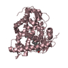
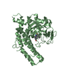
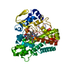
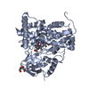
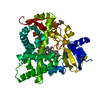
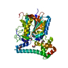
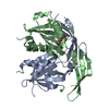
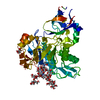
 Bacteroides thetaiotaomicron VPI-5482 (bacteria)
 X-RAY DIFFRACTION /
  Authors
 Citation
 Structure visualization
 Downloads & links
 Other downloads

 PDBj
PDBj


 Assembly
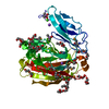


 Mass: 40.078 Da / Num. of mol.: 2 / Source method: obtained synthetically / Formula: Ca
 Mass: 22.990 Da / Num. of mol.: 2 / Source method: obtained synthetically / Formula: Na
 Mass: 136.989 Da / Num. of mol.: 2 / Source method: obtained synthetically / Formula: C2H6AsO2
 Mass: 59.044 Da / Num. of mol.: 2 / Source method: obtained synthetically / Formula: C2H3O2
 Mass: 106.120 Da / Num. of mol.: 12 / Source method: obtained synthetically / Formula: C4H10O3
 Mass: 282.331 Da / Num. of mol.: 7 / Source method: obtained synthetically / Formula: C12H26O7 / Comment: precipitant*YM
 Sample preparation
 / Beamline: BL11-1 / Wavelength: 0.97852
  Processing