 Movie
Movie Controller


+ Open data

- Basic information
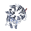
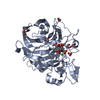
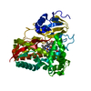
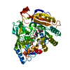
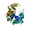
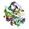
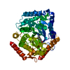
| Entry | Database: PDB / ID: 1s6c | ||||||
|---|---|---|---|---|---|---|---|
| Title | Crystal structure of the complex between KChIP1 and Kv4.2 N1-30 | ||||||
 Components Components |
| ||||||
 Keywords Keywords | TRANSPORT PROTEIN / EF-hand | ||||||
| Function / homology |  Function and homology information Function and homology informationPhase 1 - inactivation of fast Na+ channels / Kv4.3-KChIP1 channel complex / Kv4.2-KChIP2 channel complex / cardiac muscle cell action potential / A-type (transient outward) potassium channel activity / Voltage gated Potassium channels / voltage-gated monoatomic ion channel activity involved in regulation of postsynaptic membrane potential / potassium channel complex / perinuclear endoplasmic reticulum / positive regulation of action potential ...Phase 1 - inactivation of fast Na+ channels / Kv4.3-KChIP1 channel complex / Kv4.2-KChIP2 channel complex / cardiac muscle cell action potential / A-type (transient outward) potassium channel activity / Voltage gated Potassium channels / voltage-gated monoatomic ion channel activity involved in regulation of postsynaptic membrane potential / potassium channel complex / perinuclear endoplasmic reticulum / positive regulation of action potential / membrane repolarization / regulation of potassium ion transmembrane transport / anchoring junction / postsynaptic specialization membrane / locomotor rhythm / neuronal cell body membrane / plasma membrane raft / action potential / regulation of signal transduction / potassium channel regulator activity / monoatomic ion channel activity / voltage-gated potassium channel activity / potassium channel activity / neuronal action potential / voltage-gated potassium channel complex / T-tubule / potassium ion transmembrane transport / sensory perception of pain / muscle contraction / potassium ion transport / cellular response to mechanical stimulus / protein homooligomerization / sarcolemma / caveola / cellular response to xenobiotic stimulus / GABA-ergic synapse / cytoplasmic side of plasma membrane / cellular response to hypoxia / dendritic spine / transmembrane transporter binding / perikaryon / postsynaptic membrane / neuronal cell body / calcium ion binding / dendrite / protein-containing complex binding / glutamatergic synapse / membrane / metal ion binding / plasma membrane / cytoplasm Similarity search - Function | ||||||
| Biological species |  | ||||||
| Method |  X-RAY DIFFRACTION / SYNCHROTRON / MOLECULAR REPLACEMENT / Resolution: 2 Å X-RAY DIFFRACTION / SYNCHROTRON / MOLECULAR REPLACEMENT / Resolution: 2 Å | ||||||
 Authors Authors | Zhou, W. / Qian, Y. / Kunjilwar, K. / Pfaffinger, P.J. / Choe, S. | ||||||
 Citation Citation | Journal: Neuron / Year: 2004 Title: Structural insights into the functional interaction of KChIP1 with Shal-type K(+) channels. Authors: Zhou, W. / Qian, Y. / Kunjilwar, K. / Pfaffinger, P.J. / Choe, S. | ||||||
| History |
|
- Structure visualization
| Structure viewer | Molecule: MolmilJmol/JSmol |
|---|
- Downloads & links
-Download
| PDBx/mmCIF format | 1s6c.cif.gz | 56.7 KB | Display | PDBx/mmCIF format |
|---|---|---|---|---|
| PDB format | pdb1s6c.ent.gz | 39.7 KB | Display | PDB format |
| PDBx/mmJSON format | 1s6c.json.gz | Tree view | PDBx/mmJSON format | |
| Others |  Other downloads Other downloads |
-Validation report
| Arichive directory | https://data.pdbj.org/pub/pdb/validation_reports/s6/1s6cftp://data.pdbj.org/pub/pdb/validation_reports/s6/1s6c | HTTPS FTP |
|---|
-Related structure data
| Related structure data | 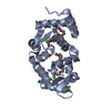 1g8iS S: Starting model for refinement |
|---|---|
| Similar structure data |


-Links
 PDBj
PDBj


- Assembly
| Deposited unit | 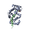
| ||||||||
|---|---|---|---|---|---|---|---|---|---|
| 1 | 
| ||||||||
| Unit cell |
|
-Components
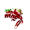
| #1: Protein | Mass: 21760.275 Da / Num. of mol.: 1 / Fragment: Core domain (Residues 34-216) Source method: isolated from a genetically manipulated source Source: (gene. exp.)  | ||||
|---|---|---|---|---|---|
| #2: Protein/peptide | Mass: 2967.552 Da / Num. of mol.: 1 / Fragment: N-terminus (Residues 1-30) Source method: isolated from a genetically manipulated source Source: (gene. exp.) | ||||
| #3: Chemical |   Mass: 40.078 Da / Num. of mol.: 2 / Source method: obtained synthetically / Formula: Ca Mass: 40.078 Da / Num. of mol.: 2 / Source method: obtained synthetically / Formula: Ca#4: Water | ChemComp-HOH / |  Mass: 18.015 Da / Num. of mol.: 150 / Source method: isolated from a natural source / Formula: H2O Mass: 18.015 Da / Num. of mol.: 150 / Source method: isolated from a natural source / Formula: H2OHas protein modification | Y | |
-Experimental details
-Experiment
| Experiment | Method: X-RAY DIFFRACTION / Number of used crystals: 1 |
|---|
- Sample preparation
| Crystal | Density Matthews: 2.4 Å3/Da / Density % sol: 48.81 % | |||||||||||||||||||||||||||||||||||||||||||||||||
|---|---|---|---|---|---|---|---|---|---|---|---|---|---|---|---|---|---|---|---|---|---|---|---|---|---|---|---|---|---|---|---|---|---|---|---|---|---|---|---|---|---|---|---|---|---|---|---|---|---|---|
| Crystal grow | Temperature: 298 K / Method: vapor diffusion, hanging drop / pH: 6 Details: PEG 1000, magnesium nitrate, sodium cacodylate, DTT, pH 6, VAPOR DIFFUSION, HANGING DROP, temperature 298K | |||||||||||||||||||||||||||||||||||||||||||||||||
| Crystal grow | *PLUS Temperature: 23 ℃ / pH: 8 / Method: vapor diffusion, hanging drop | |||||||||||||||||||||||||||||||||||||||||||||||||
| Components of the solutions | *PLUS
|
-Data collection
| Diffraction | Mean temperature: 100 K |
|---|---|
| Diffraction source | Source: SYNCHROTRON / Site: ALS  / Beamline: 8.2.1 / Wavelength: 1.044 Å / Beamline: 8.2.1 / Wavelength: 1.044 Å |
| Detector | Type: ADSC QUANTUM 210 / Detector: CCD / Date: Aug 3, 2002 |
| Radiation | Monochromator: Double Crystal Si(111) / Protocol: SINGLE WAVELENGTH / Monochromatic (M) / Laue (L): M / Scattering type: x-ray |
| Radiation wavelength | Wavelength: 1.044 Å / Relative weight: 1 |
| Reflection | Resolution: 2→100 Å / Num. all: 16062 / Num. obs: 16062 / % possible obs: 97.5 % |
| Reflection shell | Resolution: 2→2.07 Å / % possible all: 94.76 |
| Reflection | *PLUS % possible obs: 100 % / Num. measured all: 148552 / Rmerge(I) obs: 0.05 |
| Reflection shell | *PLUS % possible obs: 94.8 % / Mean I/σ(I) obs: 6 |
- Processing
| Software |
| ||||||||||||||||||||
|---|---|---|---|---|---|---|---|---|---|---|---|---|---|---|---|---|---|---|---|---|---|
| Refinement | Method to determine structure: MOLECULAR REPLACEMENT Starting model: PDB ENTRY 1G8I Resolution: 2→19.79 Å / Cross valid method: THROUGHOUT / σ(F): 0 / Stereochemistry target values: Engh & Huber
| ||||||||||||||||||||
| Refinement step | Cycle: LAST / Resolution: 2→19.79 Å
| ||||||||||||||||||||
| Refine LS restraints |
| ||||||||||||||||||||
| Refine LS restraints | *PLUS
|
