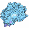
| 登録情報 | データベース: PDB / ID: 6jjy
|
|---|
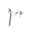
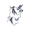
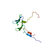
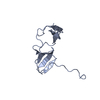
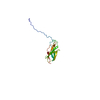
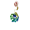
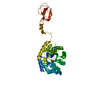
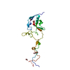
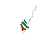
| タイトル | Crystal Structure of KIBRA and beta-Dystroglycan |
|---|
 要素 要素 | - Peptide from Dystroglycan
- Protein KIBRA
|
|---|
 キーワード キーワード | SIGNALING PROTEIN / WW tandem / PY tandem / KIBRA / PTPN14 |
|---|
| 機能・相同性 |  機能・相同性情報 機能・相同性情報
Defective POMT2 causes MDDGA2, MDDGB2 and MDDGC2 / Defective POMT1 causes MDDGA1, MDDGB1 and MDDGC1 / dystroglycan complex / nerve maturation / muscle attachment / Defective POMGNT1 causes MDDGA3, MDDGB3 and MDDGC3 / regulation of embryonic cell shape / regulation of hippo signaling / retrograde trans-synaptic signaling by trans-synaptic protein complex / positive regulation of basement membrane assembly involved in embryonic body morphogenesis ...Defective POMT2 causes MDDGA2, MDDGB2 and MDDGC2 / Defective POMT1 causes MDDGA1, MDDGB1 and MDDGC1 / dystroglycan complex / nerve maturation / muscle attachment / Defective POMGNT1 causes MDDGA3, MDDGB3 and MDDGC3 / regulation of embryonic cell shape / regulation of hippo signaling / retrograde trans-synaptic signaling by trans-synaptic protein complex / positive regulation of basement membrane assembly involved in embryonic body morphogenesis / O-linked glycosylation / Signaling by Hippo / contractile ring / regulation of gastrulation / calcium-dependent cell-matrix adhesion / microtubule anchoring / morphogenesis of an epithelial sheet / dystrophin-associated glycoprotein complex / laminin-1 binding / response to denervation involved in regulation of muscle adaptation / basement membrane organization / positive regulation of myelination / regulation of epithelial to mesenchymal transition / negative regulation of organ growth / dystroglycan binding / branching involved in salivary gland morphogenesis / nerve development / skeletal muscle tissue regeneration / cellular response to cholesterol / photoreceptor ribbon synapse / vinculin binding / EGR2 and SOX10-mediated initiation of Schwann cell myelination / myelination in peripheral nervous system / node of Ranvier / costamere / angiogenesis involved in wound healing / commissural neuron axon guidance / response to muscle activity / axon regeneration / regulation of neurotransmitter receptor localization to postsynaptic specialization membrane / structural constituent of muscle / positive regulation of cell-matrix adhesion / postsynaptic cytosol / epithelial tube branching involved in lung morphogenesis / positive regulation of oligodendrocyte differentiation / regulation of synapse organization / alpha-actinin binding / plasma membrane raft / membrane protein ectodomain proteolysis / Non-integrin membrane-ECM interactions / basement membrane / negative regulation of phosphatidylinositol 3-kinase/protein kinase B signal transduction / ECM proteoglycans / negative regulation of hippo signaling / negative regulation of MAPK cascade / heart morphogenesis / GABA-ergic synapse / laminin binding / tubulin binding / SH2 domain binding / nuclear periphery / negative regulation of cell migration / filopodium / axon guidance / morphogenesis of an epithelium / adherens junction / regulation of synaptic plasticity / kinase binding / sarcolemma / response to peptide hormone / ruffle membrane / Regulation of expression of SLITs and ROBOs / cellular response to mechanical stimulus / Golgi lumen / protein transport / cell migration / lamellipodium / virus receptor activity / actin binding / protein-macromolecule adaptor activity / basolateral plasma membrane / collagen-containing extracellular matrix / postsynaptic membrane / positive regulation of MAPK cascade / molecular adaptor activity / transcription coactivator activity / cytoskeleton / negative regulation of cell population proliferation / endoplasmic reticulum lumen / external side of plasma membrane / intracellular membrane-bounded organelle / focal adhesion / glutamatergic synapse / calcium ion binding / regulation of DNA-templated transcription / protein-containing complex binding / perinuclear region of cytoplasm / negative regulation of transcription by RNA polymerase II / protein-containing complex / extracellular space類似検索 - 分子機能 WWC, C2 domain / : / Dystroglycan-type cadherin-like / Dystroglycan, C-terminal / Alpha-dystroglycan domain 2 / DG-type SEA domain / Alpha-dystroglycan N-terminal domain 2 / Dystroglycan (Dystrophin-associated glycoprotein 1) / Alpha-Dystroglycan N-terminal domain 2 / DG-type SEA domain profile. ...WWC, C2 domain / : / Dystroglycan-type cadherin-like / Dystroglycan, C-terminal / Alpha-dystroglycan domain 2 / DG-type SEA domain / Alpha-dystroglycan N-terminal domain 2 / Dystroglycan (Dystrophin-associated glycoprotein 1) / Alpha-Dystroglycan N-terminal domain 2 / DG-type SEA domain profile. / Dystroglycan-type cadherin-like domains. / Putative Ig domain / Cadherin-like superfamily / C2 domain / WW domain / WW/rsp5/WWP domain signature. / C2 domain / C2 domain profile. / WW domain superfamily / WW/rsp5/WWP domain profile. / Domain with 2 conserved Trp (W) residues / WW domain / C2 domain superfamily / Immunoglobulin-like fold類似検索 - ドメイン・相同性 |
|---|
| 生物種 |   Mus musculus (ハツカネズミ) Mus musculus (ハツカネズミ)
 Homo sapiens (ヒト) Homo sapiens (ヒト) |
|---|
| 手法 |  X線回折 / X線回折 /  シンクロトロン / シンクロトロン /  分子置換 / 解像度: 2.298 Å 分子置換 / 解像度: 2.298 Å |
|---|
 データ登録者 データ登録者 | Lin, Z. / Yang, Z. / Ji, Z. / Zhang, M. |
|---|
 引用 引用 |  ジャーナル: Elife / 年: 2019 ジャーナル: Elife / 年: 2019
タイトル: Decoding WW domain tandem-mediated target recognitions in tissue growth and cell polarity.
著者: Lin, Z. / Yang, Z. / Xie, R. / Ji, Z. / Guan, K. / Zhang, M. |
|---|
| 履歴 | | 登録 | 2019年2月27日 | 登録サイト: PDBJ / 処理サイト: PDBJ |
|---|
| 改定 1.0 | 2019年9月25日 | Provider: repository / タイプ: Initial release |
|---|
| 改定 1.1 | 2023年11月22日 | Group: Data collection / Database references / Refinement description
カテゴリ: chem_comp_atom / chem_comp_bond ...chem_comp_atom / chem_comp_bond / database_2 / pdbx_initial_refinement_model
Item: _database_2.pdbx_DOI / _database_2.pdbx_database_accession |
|---|
|
|---|
 データを開く
 基本情報
基本情報 要素
要素 キーワード
キーワード 機能・相同性情報
 Homo sapiens (ヒト)
 X線回折 /
  シンクロトロン /
  分子置換 / 解像度: 2.298 Å
  データ登録者
 引用
引用 ジャーナル: Elife / 年: 2019
 構造の表示
構造の表示 Molmil
 Jmol/JSmol
 ダウンロードとリンク
 ダウンロード
 6jjy.cif.gz
 PDBx/mmCIF形式
 pdb6jjy.ent.gz
 PDB形式
PDB形式 6jjy.json.gz
 PDBx/mmJSON形式
 その他のダウンロード
 6jjy_validation.pdf.gz
 wwPDB検証レポート
 6jjy_full_validation.pdf.gz
 6jjy_validation.xml.gz
 6jjy_validation.cif.gz
 https://data.pdbj.org/pub/pdb/validation_reports/jj/6jjy
 ftp://data.pdbj.org/pub/pdb/validation_reports/jj/6jjy
 リンク
リンク 集合体
集合体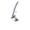
 要素
要素

 Homo sapiens (ヒト) / 遺伝子: DAG1 / 発現宿主:
 X線回折 / 使用した結晶の数: 1
  試料調製
試料調製 シンクロトロン / サイト:
  SSRF
SSRF  / ビームライン: BL19U1 / 波長: 0.9791 Å
 解析
解析 分子置換
分子置換 ムービー
ムービー コントローラー





 PDBj
PDBj


