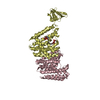
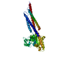
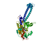
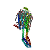
Journal: J Mol Biol / Year: 2006 Title: Evolution of bacteriophage tails: Structure of T4 gene product 10. Authors: Petr G Leiman / Mikhail M Shneider / Vadim V Mesyanzhinov / Michael G Rossmann / Abstract: The success of tailed bacteriophages to infect cells far exceeds that of most other viruses on account of their specialized tail and associated baseplate structures. The baseplate protein gene ...The success of tailed bacteriophages to infect cells far exceeds that of most other viruses on account of their specialized tail and associated baseplate structures. The baseplate protein gene product (gp) 10 of bacteriophage T4, whose structure was determined to 1.2 A resolution, was fitted into the cryo-electron microscopy structures of the pre and post-infection conformations of the virus. gp10 functions as a molecular lever that rotates and extends the hinged short tail fibers to facilitate cell attachment. The central folding motif of the gp10 trimer is similar to that of the baseplate protein gp11 and to the receptor-binding domain of the short tail fiber, gp12. The three proteins comprise the periphery of the baseplate and interact with each other. The structural and functional similarities of gp10, gp11, and gp12 and their sequential order in the T4 genome suggest that they evolved separately, subsequent to gene triplication from a common ancestor. Such events are usual in the evolution of complex organelles from a common primordial molecule.
History
Deposition
Jan 4, 2006
Deposition site: RCSB / Processing site: RCSB
Revision 1.0
Apr 4, 2006
Provider: repository / Type: Initial release
Revision 1.1
May 1, 2008
Group: Version format compliance
Revision 1.2
Jul 13, 2011
Group: Derived calculations / Version format compliance
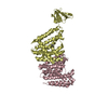
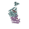
The biological assembly is a trimer. The second and third chains can be generated by using the symmetry operators of the crystallographic threefold axis: -y+1, x-y, z and y-x+1, -x+1, z
-
Components
-
Protein , 1 types, 1 molecules A
#1: Protein
BaseplatestructuralproteinGp10 / Baseplate wedge protein 10
Mass: 22432.246 Da / Num. of mol.: 1 / Fragment: C-terminal domain / Mutation: A442E, A530T Source method: isolated from a genetically manipulated source Source: (gene. exp.) Enterobacteria phage T4 (virus) / Genus: T4-like viruses / Species: Enterobacteria phage T4 sensu lato / Strain: D / Gene: 10 / Plasmid: pET28a / Species (production host): Escherichia coli / Production host: Escherichia coli BL21(DE3) (bacteria) / Strain (production host): BL21/DE3 / References: UniProt: P10928
In the structure databanks used in Yorodumi, some data are registered as the other names, "COVID-19 virus" and "2019-nCoV". Here are the details of the virus and the list of structure data.
Jan 31, 2019. EMDB accession codes are about to change! (news from PDBe EMDB page)
EMDB accession codes are about to change! (news from PDBe EMDB page)
The allocation of 4 digits for EMDB accession codes will soon come to an end. Whilst these codes will remain in use, new EMDB accession codes will include an additional digit and will expand incrementally as the available range of codes is exhausted. The current 4-digit format prefixed with “EMD-” (i.e. EMD-XXXX) will advance to a 5-digit format (i.e. EMD-XXXXX), and so on. It is currently estimated that the 4-digit codes will be depleted around Spring 2019, at which point the 5-digit format will come into force.
The EM Navigator/Yorodumi systems omit the EMD- prefix.
Related info.:Q: What is EMD? / ID/Accession-code notation in Yorodumi/EM Navigator
Yorodumi is a browser for structure data from EMDB, PDB, SASBDB, etc.
This page is also the successor to EM Navigator detail page, and also detail information page/front-end page for Omokage search.
The word "yorodu" (or yorozu) is an old Japanese word meaning "ten thousand". "mi" (miru) is to see.
Related info.:EMDB / PDB / SASBDB / Comparison of 3 databanks / Yorodumi Search / Aug 31, 2016. New EM Navigator & Yorodumi / Yorodumi Papers / Jmol/JSmol / Function and homology information / Changes in new EM Navigator and Yorodumi
 Movie
Movie Controller

 Yorodumi
 Open data
 Basic information
 Components
 Keywords
 Function and homology information
 Enterobacteria phage T4 (virus)
 X-RAY DIFFRACTION /
  Authors
 Citation
 Structure visualization
 Downloads & links
 Other downloads


 PDBj
PDBj

 Assembly
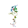


 Mass: 79.904 Da / Num. of mol.: 1 / Source method: obtained synthetically / Formula: Br
 Mass: 78.972 Da / Num. of mol.: 1 / Source method: obtained synthetically / Formula: PO3
 Mass: 122.143 Da / Num. of mol.: 1 / Source method: obtained synthetically / Formula: C4H12NO3 / Comment: pH buffer*YM
 Mass: 62.068 Da / Num. of mol.: 4 / Source method: obtained synthetically / Formula: C2H6O2
 Sample preparation
 Processing