 Movie
Movie Controller

[English] 日本語
 Yorodumi
- PDB-2fl9: Evolution of bacteriophage tails: Structure of T4 gene product 10 -
+ Open data

- Basic information
| Entry | Database: PDB / ID: 2fl9 | ||||||
|---|---|---|---|---|---|---|---|
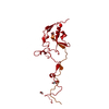
| Title | Evolution of bacteriophage tails: Structure of T4 gene product 10 | ||||||
 Components Components | Baseplate structural protein Gp10 | ||||||
 Keywords Keywords | VIRUS/VIRAL PROTEIN / Bacteriophage T4 / Baseplate / Tail / Evolution / gp10 / Structural comparisons / VIRUS-VIRAL PROTEIN COMPLEX | ||||||
| Function / homology |  Function and homology information Function and homology informationvirus tail, baseplate / viral tail assembly / viral release from host cell Similarity search - Function | ||||||
| Biological species |  Enterobacteria phage T4 (virus) Enterobacteria phage T4 (virus) | ||||||
| Method | ELECTRON MICROSCOPY / single particle reconstruction / cryo EM / Resolution: 17 Å | ||||||
 Authors Authors | Leiman, P.G. / Shneider, M.M. / Mesyanzhinov, V.V. / Rossmann, M.G. | ||||||
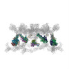
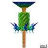
 Citation Citation | Journal: J Mol Biol / Year: 2006 Title: Evolution of bacteriophage tails: Structure of T4 gene product 10. Authors: Petr G Leiman / Mikhail M Shneider / Vadim V Mesyanzhinov / Michael G Rossmann /  Abstract: The success of tailed bacteriophages to infect cells far exceeds that of most other viruses on account of their specialized tail and associated baseplate structures. The baseplate protein gene ...The success of tailed bacteriophages to infect cells far exceeds that of most other viruses on account of their specialized tail and associated baseplate structures. The baseplate protein gene product (gp) 10 of bacteriophage T4, whose structure was determined to 1.2 A resolution, was fitted into the cryo-electron microscopy structures of the pre and post-infection conformations of the virus. gp10 functions as a molecular lever that rotates and extends the hinged short tail fibers to facilitate cell attachment. The central folding motif of the gp10 trimer is similar to that of the baseplate protein gp11 and to the receptor-binding domain of the short tail fiber, gp12. The three proteins comprise the periphery of the baseplate and interact with each other. The structural and functional similarities of gp10, gp11, and gp12 and their sequential order in the T4 genome suggest that they evolved separately, subsequent to gene triplication from a common ancestor. Such events are usual in the evolution of complex organelles from a common primordial molecule. | ||||||
| History |
|
- Structure visualization
| Movie |
Movie viewer |
|---|---|
| Structure viewer | Molecule: MolmilJmol/JSmol |
- Downloads & links
-Download
| PDBx/mmCIF format | 2fl9.cif.gz | 234.3 KB | Display | PDBx/mmCIF format |
|---|---|---|---|---|
| PDB format | pdb2fl9.ent.gz | 135.2 KB | Display | PDB format |
| PDBx/mmJSON format | 2fl9.json.gz | Tree view | PDBx/mmJSON format | |
| Others |  Other downloads Other downloads |
-Validation report
| Arichive directory | https://data.pdbj.org/pub/pdb/validation_reports/fl/2fl9ftp://data.pdbj.org/pub/pdb/validation_reports/fl/2fl9 | HTTPS FTP |
|---|
-Related structure data


-Links
 PDBj
PDBj- Assembly
| Deposited unit | 
|
|---|---|
| 1 |
|
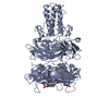
| Symmetry | Point symmetry: (Schoenflies symbol: C6 (6 fold cyclic)) |
-Components
| #1: Protein | Mass: 66369.742 Da / Num. of mol.: 18 / Mutation: A442E, A530T Source method: isolated from a genetically manipulated source Source: (gene. exp.) Enterobacteria phage T4 (virus) / Genus: T4-like viruses / Species: Enterobacteria phage T4 sensu lato / Strain: D / Gene: 10 / Species (production host): Escherichia coli / Production host:  |
|---|
-Experimental details
-Experiment
| Experiment | Method: ELECTRON MICROSCOPY |
|---|---|
| EM experiment | Aggregation state: PARTICLE / 3D reconstruction method: single particle reconstruction |
- Sample preparation
| Component | Name: bacteriophage T4 baseplate / Type: VIRUS Details: The trimeric gene product 10 is a structural component of the sixfold-symmetric T4 baseplate. The entry contains the fit of the gp10 C-terminal domain crystal structure (residues 406-602) ...Details: The trimeric gene product 10 is a structural component of the sixfold-symmetric T4 baseplate. The entry contains the fit of the gp10 C-terminal domain crystal structure (residues 406-602) and the homology model of the gp10 N-terminal domain (residues 1-160) into the cryoEM map of the T4 baseplate in the star conformation (EMDB ID 1086). The homology model of the N-terminal domains is based on the crystal structure of T4 gp9 (PDB ID 1S2E). Source: NATURAL |
|---|---|
| Source (natural) | Organism: Enterobacteria phage T4 (virus) |
| Details of virus | Host category: BACTERIA / Isolate: STRAIN / Type: VIRION |
| Natural host | Organism: Escherichia coli |
| Buffer solution | pH: 7.5 / Details: H2O |
| Specimen | Embedding applied: NO / Shadowing applied: NO / Staining applied: NO / Vitrification applied: YES |
| Vitrification | Cryogen name: ETHANE |
- Electron microscopy imaging
| Microscopy | Model: FEI/PHILIPS CM300FEG/T / Date: Jan 6, 2002 |
|---|---|
| Electron gun | Electron source:  FIELD EMISSION GUN / Accelerating voltage: 300 kV / Illumination mode: SPOT SCAN FIELD EMISSION GUN / Accelerating voltage: 300 kV / Illumination mode: SPOT SCAN |
| Electron lens | Mode: BRIGHT FIELD / Nominal magnification: 45000 X / Nominal defocus max: 3400 nm / Nominal defocus min: 500 nm |
| Image recording | Electron dose: 20 e/Å2 / Film or detector model: KODAK SO-163 FILM |
- Processing
| EM software |
| |||||||||||||||||||||
|---|---|---|---|---|---|---|---|---|---|---|---|---|---|---|---|---|---|---|---|---|---|---|
| Symmetry | Point symmetry: C6 (6 fold cyclic) | |||||||||||||||||||||
| 3D reconstruction | Resolution: 17 Å / Num. of particles: 1965 Details: THE COORDINATES CONTAIN CA ONLY. RESIDUES 161-405 ARE MISSING. PLEASE SEE THE RELATED EMDB ENTRY FOR DETAILS OF THE EM RECONSTRUCTION Symmetry type: POINT | |||||||||||||||||||||
| Atomic model building | Protocol: RIGID BODY FIT / Space: REAL Target criteria: correlation coefficient as defined by the program COLORES/SITUS Details: METHOD--laplacian filtered real space REFINEMENT PROTOCOL--rigid body | |||||||||||||||||||||
| Atomic model building |
| |||||||||||||||||||||
| Refinement step | Cycle: LAST
|
