[English] 日本語
Yorodumi
- EMDB-1089: Three-dimensional rearrangement of proteins in the tail of bacter... -

+
Open data


ID or keywords:

Loading...

-
Basic information

Entry
Database: EMDB / ID: EMD-1089
TitleThree-dimensional rearrangement of proteins in the tail of bacteriophage T4 on infection of its host.
Map dataBacteriophage T4 contracted tail. This reconstruction has been calculated using the same sample and micrographs as EMD-1086 but in a larger box size
Sample
  • Sample: T4 phages treated with 3 M urea
  • Virus: Enterobacteria phage T4 (virus)
Function / homology
Function and homology information


viral tail assembly / virus tail, baseplate / viral release from host cell
Similarity search - Function
Baseplate structural protein Gp11 / Bacteriophage T4, Gp11, C-terminal finger domain / Baseplate structural protein Gp11, N-terminal domain superfamily / Baseplate structural protein Gp11 superfamily / Baseplate structural protein Gp11, C-terminal domain / GP11 baseplate wedge protein / Baseplate structural protein Gp9 C-terminal domain superfamily / Bacteriophage T4, Gp8 / Bacteriophage T4, Gp8 superfamily / Bacteriophage T4, Gp8 ...Baseplate structural protein Gp11 / Bacteriophage T4, Gp11, C-terminal finger domain / Baseplate structural protein Gp11, N-terminal domain superfamily / Baseplate structural protein Gp11 superfamily / Baseplate structural protein Gp11, C-terminal domain / GP11 baseplate wedge protein / Baseplate structural protein Gp9 C-terminal domain superfamily / Bacteriophage T4, Gp8 / Bacteriophage T4, Gp8 superfamily / Bacteriophage T4, Gp8 / Baseplate structural protein Gp9/Gp10 / Baseplate structural protein Gp9/Gp10 middle domain superfamily / Gp9-like superfamily / Bacteriophage T4 gp9/10-like protein
Similarity search - Domain/homology
Baseplate protein gp9 / Baseplate wedge protein gp11 / Baseplate wedge protein gp8
Similarity search - Component
Biological speciesEnterobacteria phage T4 (virus)
Methodsingle particle reconstruction / cryo EM / Resolution: 18.0 Å
AuthorsLeiman PG / Chipman PR / Kostyuchenko VA / Mesyanzhinov VV / Rossmann MG
Citation
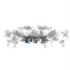
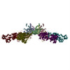
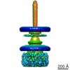
Journal: Cell / Year: 2004
Title: Three-dimensional rearrangement of proteins in the tail of bacteriophage T4 on infection of its host.
Authors: Petr G Leiman / Paul R Chipman / Victor A Kostyuchenko / Vadim V Mesyanzhinov / Michael G Rossmann /
Abstract: The contractile tail of bacteriophage T4 undergoes major structural transitions when the virus attaches to the host cell surface. The baseplate at the distal end of the tail changes from a hexagonal ...The contractile tail of bacteriophage T4 undergoes major structural transitions when the virus attaches to the host cell surface. The baseplate at the distal end of the tail changes from a hexagonal to a star shape. This causes the sheath around the tail tube to contract and the tail tube to protrude from the baseplate and pierce the outer cell membrane and the cell wall before reaching the inner cell membrane for subsequent viral DNA injection. Analogously, the T4 tail can be contracted by treatment with 3 M urea. The structure of the T4 contracted tail, including the head-tail joining region, has been determined by cryo-electron microscopy to 17 A resolution. This 1200 A-long, 20 MDa structure has been interpreted in terms of multiple copies of its approximately 20 component proteins. A comparison with the metastable hexagonal baseplate of the mature virus shows that the baseplate proteins move as rigid bodies relative to each other during the structural change.
History
DepositionJul 19, 2004-
Header (metadata) releaseJul 19, 2004-
Map releaseSep 7, 2004-
UpdateMay 26, 2011-
Current statusMay 26, 2011Processing site: PDBe / Status: Released

-
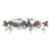
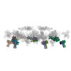
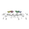
Structure visualization

Movie
  • Surface view with section colored by density value
  • Surface level: 2
  • Imaged by UCSF Chimera
  • Download
  • Surface view colored by cylindrical radius
  • Surface level: 2
  • Imaged by UCSF Chimera
  • Download
  • Surface view with fitted model
  • Atomic models: PDB-3j2n
  • Surface level: 2
  • Imaged by UCSF Chimera
  • Download
Movie viewer
Structure viewerEM map:
SurfViewMolmilJmol/JSmol
Supplemental images

Downloads & links

-
Map

FileDownload / File: emd_1089.map.gz / Format: CCP4 / Size: 57.4 MB / Type: IMAGE STORED AS FLOATING POINT NUMBER (4 BYTES)
AnnotationBacteriophage T4 contracted tail. This reconstruction has been calculated using the same sample and micrographs as EMD-1086 but in a larger box size
Voxel sizeX=Y=Z: 4.51104 Å
Density
Contour Level1: 1.5 / Movie #1: 2
Minimum - Maximum-11.4809 - 15.4794
Average (Standard dev.)0.000000000138942 (±1.0)
SymmetrySpace group: 1
Details

EMDB XML:

Map geometry
Axis orderXYZ
Origin-111-111-190
Dimensions223223310
Spacing223223310
CellA: 1005.96 Å / B: 1005.96 Å / C: 1398.42 Å
α=β=γ: 90 °

CCP4 map header:

modeImage stored as Reals
Å/pix. X/Y/Z4.51104035874444.51104035874444.5110387096774
M x/y/z223223310
origin x/y/z0.0000.0000.000
length x/y/z1005.9621005.9621398.422
α/β/γ90.00090.00090.000
start NX/NY/NZ-96-56-288
NX/NY/NZ192112576
MAP C/R/S123
start NC/NR/NS-111-111-190
NC/NR/NS223223310
D min/max/mean-11.48115.4790.000

-
Supplemental data

-
Sample components

-
Entire : T4 phages treated with 3 M urea

EntireName: T4 phages treated with 3 M urea
Components
  • Sample: T4 phages treated with 3 M urea
  • Virus: Enterobacteria phage T4 (virus)

-
Supramolecule #1000: T4 phages treated with 3 M urea

SupramoleculeName: T4 phages treated with 3 M urea / type: sample / ID: 1000 / Number unique components: 1
Molecular weightExperimental: 220 MDa / Theoretical: 220 MDa / Method: Estimate

-
Supramolecule #1: Enterobacteria phage T4

SupramoleculeName: Enterobacteria phage T4 / type: virus / ID: 1 / Name.synonym: phage T4 / Details: treated with 3 M urea / NCBI-ID: 10665 / Sci species name: Enterobacteria phage T4 / Virus type: VIRION / Virus isolate: STRAIN / Virus enveloped: No / Virus empty: No / Syn species name: phage T4
Host (natural)Organism: Escherichia coli (E. coli) / synonym: BACTERIA(EUBACTERIA)
Molecular weightExperimental: 220 MDa / Theoretical: 220 MDa

-
Experimental details

-
Structure determination

Methodcryo EM
Processingsingle particle reconstruction
Aggregation stateparticle

-
Sample preparation

Concentration5 mg/mL
BufferpH: 7.5 / Details: H2O
GridDetails: 200 mesh cupper grid
VitrificationCryogen name: ETHANE / Chamber temperature: 100 K

-
Electron microscopy

MicroscopeFEI/PHILIPS CM300FEG/T
Electron beamAcceleration voltage: 300 kV / Electron source: FIELD EMISSION GUN
Electron opticsCalibrated magnification: 47000 / Illumination mode: SPOT SCAN / Imaging mode: BRIGHT FIELDBright-field microscopy / Nominal defocus max: 3.4 µm / Nominal defocus min: 0.5 µm / Nominal magnification: 45000
Specialist opticsEnergy filter - Name: FEI
Sample stageSpecimen holder: Side entry liquid nitrogen-cooled cryo specimen holder
Specimen holder model: GATAN LIQUID NITROGEN
TemperatureAverage: 100 K
DateJan 6, 2002
Image recordingCategory: FILM / Film or detector model: KODAK SO-163 FILM / Digitization - Scanner: ZEISS SCAI / Digitization - Sampling interval: 4.51104 µm / Number real images: 100 / Average electron dose: 20 e/Å2 / Bits/pixel: 8
Tilt angle min0
Tilt angle max0

-
Image processing

CTF correctionDetails: Each particle
Final angle assignmentDetails: theta 45 degrees, phi 180 degrees
Final reconstructionApplied symmetry - Point group: C6 (6 fold cyclic) / Algorithm: OTHER / Resolution.type: BY AUTHOR / Resolution: 18.0 Å / Resolution method: FSC 0.5 CUT-OFF / Software - Name: Spider / Number images used: 1965

-
Atomic model buiding 1

DetailsThe components were separately fitted using Situs v.2.0.
Output model

PDB-1tja:
Fitting of gp8, gp9, and gp11 into the cryo-EM reconstruction of the bacteriophage T4 contracted tail

+
About Yorodumi

-
News

-
Feb 9, 2022. New format data for meta-information of EMDB entries

New format data for meta-information of EMDB entries

  • Version 3 of the EMDB header file is now the official format.
  • The previous official version 1.9 will be removed from the archive.

Related info.:EMDB header

External links:wwPDB to switch to version 3 of the EMDB data model

-
Aug 12, 2020. Covid-19 info

Covid-19 info

URL: https://pdbj.org/emnavi/covid19.php

New page: Covid-19 featured information page in EM Navigator.

Related info.:Covid-19 info / Mar 5, 2020. Novel coronavirus structure data

+
Mar 5, 2020. Novel coronavirus structure data

Novel coronavirus structure data

Related info.:Yorodumi Speices / Aug 12, 2020. Covid-19 info

External links:COVID-19 featured content - PDBj / Molecule of the Month (242):Coronavirus Proteases

+
Jan 31, 2019. EMDB accession codes are about to change! (news from PDBe EMDB page)

EMDB accession codes are about to change! (news from PDBe EMDB page)

  • The allocation of 4 digits for EMDB accession codes will soon come to an end. Whilst these codes will remain in use, new EMDB accession codes will include an additional digit and will expand incrementally as the available range of codes is exhausted. The current 4-digit format prefixed with “EMD-” (i.e. EMD-XXXX) will advance to a 5-digit format (i.e. EMD-XXXXX), and so on. It is currently estimated that the 4-digit codes will be depleted around Spring 2019, at which point the 5-digit format will come into force.
  • The EM Navigator/Yorodumi systems omit the EMD- prefix.

Related info.:Q: What is EMD? / ID/Accession-code notation in Yorodumi/EM Navigator

External links:EMDB Accession Codes are Changing Soon! / Contact to PDBj

+
Jul 12, 2017. Major update of PDB

Major update of PDB

  • wwPDB released updated PDB data conforming to the new PDBx/mmCIF dictionary.
  • This is a major update changing the version number from 4 to 5, and with Remediation, in which all the entries are updated.
  • In this update, many items about electron microscopy experimental information are reorganized (e.g. em_software).
  • Now, EM Navigator and Yorodumi are based on the updated data.

External links:wwPDB Remediation / Enriched Model Files Conforming to OneDep Data Standards Now Available in the PDB FTP Archive

-
Yorodumi

Thousand views of thousand structures

  • Yorodumi is a browser for structure data from EMDB, PDB, SASBDB, etc.
  • This page is also the successor to EM Navigator detail page, and also detail information page/front-end page for Omokage search.
  • The word "yorodu" (or yorozu) is an old Japanese word meaning "ten thousand". "mi" (miru) is to see.

Related info.:EMDB / PDB / SASBDB / Comparison of 3 databanks / Yorodumi Search / Aug 31, 2016. New EM Navigator & Yorodumi / Yorodumi Papers / Jmol/JSmol / Function and homology information / Changes in new EM Navigator and Yorodumi

Read more