 Movie
Movie Controller

 Yorodumi
+ Open data

- Basic information
| Entry | Database: EMDB / ID: EMD-1086 | |||||||||
|---|---|---|---|---|---|---|---|---|---|---|
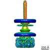
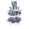
| Title | Three-dimensional rearrangement of proteins in the tail of bacteriophage T4 on infection of its host. | |||||||||
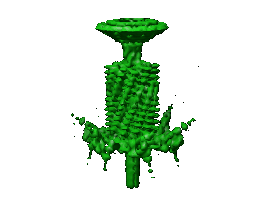
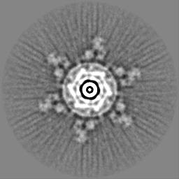
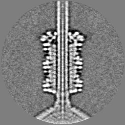
 Map data Map data | CryoEM density of the T4 tail with contracted sheath and the baseplate in the star conformation. | |||||||||
 Sample Sample |
| |||||||||
| Function / homology |  Function and homology information Function and homology informationvirus tail, sheath / symbiont genome ejection through host cell envelope, contractile tail mechanism / virus tail, baseplate / viral tail assembly / viral release from host cell / virion component / identical protein binding Similarity search - Function | |||||||||
| Biological species |  Enterobacteria phage T4 (virus) Enterobacteria phage T4 (virus) | |||||||||
| Method | single particle reconstruction / cryo EM / Resolution: 16.0 Å | |||||||||
 Authors Authors | Leiman PG / Chipman PR / Kostyuchenko VA / Mesyanzhinov VV / Rossmann MG | |||||||||
 Citation Citation | Journal: Cell / Year: 2004 Title: Three-dimensional rearrangement of proteins in the tail of bacteriophage T4 on infection of its host. Authors: Petr G Leiman / Paul R Chipman / Victor A Kostyuchenko / Vadim V Mesyanzhinov / Michael G Rossmann /  Abstract: The contractile tail of bacteriophage T4 undergoes major structural transitions when the virus attaches to the host cell surface. The baseplate at the distal end of the tail changes from a hexagonal ...The contractile tail of bacteriophage T4 undergoes major structural transitions when the virus attaches to the host cell surface. The baseplate at the distal end of the tail changes from a hexagonal to a star shape. This causes the sheath around the tail tube to contract and the tail tube to protrude from the baseplate and pierce the outer cell membrane and the cell wall before reaching the inner cell membrane for subsequent viral DNA injection. Analogously, the T4 tail can be contracted by treatment with 3 M urea. The structure of the T4 contracted tail, including the head-tail joining region, has been determined by cryo-electron microscopy to 17 A resolution. This 1200 A-long, 20 MDa structure has been interpreted in terms of multiple copies of its approximately 20 component proteins. A comparison with the metastable hexagonal baseplate of the mature virus shows that the baseplate proteins move as rigid bodies relative to each other during the structural change. | |||||||||
| History |
|
- Structure visualization
| Movie |
Movie viewer |
|---|---|
| Structure viewer | EM map: SurfViewMolmilJmol/JSmol |
| Supplemental images |
- Downloads & links
-EMDB archive
| Map data | emd_1086.map.gz | 7.8 MB | EMDB map data format | |
|---|---|---|---|---|
| Header (meta data) | emd-1086-v30.xmlemd-1086.xml | 11.5 KB 11.5 KB | Display Display | EMDB header |
| Images |  1086.gif 1086.gif | 11.9 KB | ||
| Archive directory |  http://ftp.pdbj.org/pub/emdb/structures/EMD-1086ftp://ftp.pdbj.org/pub/emdb/structures/EMD-1086 http://ftp.pdbj.org/pub/emdb/structures/EMD-1086ftp://ftp.pdbj.org/pub/emdb/structures/EMD-1086 | HTTPS FTP |
-Related structure data
| Related structure data | 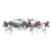 1tjaMC  2fl9M  3foiM 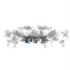 3h3yM 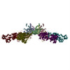 3j2nM 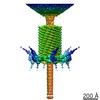 1089C M: atomic model generated by this map C: citing same article ( |
|---|---|
| Similar structure data |
-Links
| EMDB pages | EMDB (EBI/PDBe) / EMDataResource |
|---|
-Map
| File | Download / File: emd_1086.map.gz / Format: CCP4 / Size: 62.5 MB / Type: IMAGE STORED AS FLOATING POINT NUMBER (4 BYTES) | ||||||||||||||||||||||||||||||||||||||||||||||||||||||||||||||||||||
|---|---|---|---|---|---|---|---|---|---|---|---|---|---|---|---|---|---|---|---|---|---|---|---|---|---|---|---|---|---|---|---|---|---|---|---|---|---|---|---|---|---|---|---|---|---|---|---|---|---|---|---|---|---|---|---|---|---|---|---|---|---|---|---|---|---|---|---|---|---|
| Annotation | CryoEM density of the T4 tail with contracted sheath and the baseplate in the star conformation. | ||||||||||||||||||||||||||||||||||||||||||||||||||||||||||||||||||||
| Projections & slices | Image control
Images are generated by Spider. | ||||||||||||||||||||||||||||||||||||||||||||||||||||||||||||||||||||
| Voxel size | X=Y=Z: 3.93285 Å | ||||||||||||||||||||||||||||||||||||||||||||||||||||||||||||||||||||
| Density |
| ||||||||||||||||||||||||||||||||||||||||||||||||||||||||||||||||||||
| Symmetry | Space group: 1 | ||||||||||||||||||||||||||||||||||||||||||||||||||||||||||||||||||||
| Details | EMDB XML:
CCP4 map header:
| ||||||||||||||||||||||||||||||||||||||||||||||||||||||||||||||||||||
 Z (Sec.)
 Y (Row.)
 X (Col.)


-Supplemental data
- Sample components
-Entire : T4 phages treated with 3 M urea
| Entire | Name: T4 phages treated with 3 M urea |
|---|---|
| Components |
|
-Supramolecule #1000: T4 phages treated with 3 M urea
| Supramolecule | Name: T4 phages treated with 3 M urea / type: sample / ID: 1000 / Number unique components: 1 |
|---|---|
| Molecular weight | Experimental: 220 MDa / Theoretical: 220 MDa / Method: Estimate |
-Supramolecule #1: Enterobacteria phage T4
| Supramolecule | Name: Enterobacteria phage T4 / type: virus / ID: 1 / Name.synonym: phage T4 / Details: treated with 3 M urea / NCBI-ID: 10665 / Sci species name: Enterobacteria phage T4 / Virus type: VIRION / Virus isolate: STRAIN / Virus enveloped: No / Virus empty: No / Syn species name: phage T4 |
|---|---|
| Host (natural) | Organism:  |
| Molecular weight | Experimental: 220 MDa / Theoretical: 220 MDa |
-Experimental details
-Structure determination
| Method | cryo EM |
|---|---|
 Processing Processing | single particle reconstruction |
| Aggregation state | particle |
-Sample preparation
| Concentration | 5 mg/mL |
|---|---|
| Buffer | pH: 7.5 / Details: H2O |
| Grid | Details: 200 mesh cupper grid |
| Vitrification | Cryogen name: ETHANE / Chamber temperature: 100 K |
- Electron microscopy
| Microscope | FEI/PHILIPS CM300FEG/T |
|---|---|
| Temperature | Average: 100 K |
| Specialist optics | Energy filter - Name: FEI |
| Date | Jan 6, 2002 |
| Image recording | Category: FILM / Film or detector model: KODAK SO-163 FILM / Digitization - Scanner: ZEISS SCAI / Digitization - Sampling interval: 3.93285 µm / Number real images: 100 / Average electron dose: 20 e/Å2 / Bits/pixel: 8 |
| Tilt angle min | 0 |
| Tilt angle max | 0 |
| Electron beam | Acceleration voltage: 300 kV / Electron source:  FIELD EMISSION GUN FIELD EMISSION GUN |
| Electron optics | Calibrated magnification: 47000 / Illumination mode: SPOT SCAN / Imaging mode: BRIGHT FIELD / Nominal defocus max: 3.4 µm / Nominal defocus min: 0.5 µm / Nominal magnification: 45000 |
| Sample stage | Specimen holder: Side entry liquid nitrogen-cooled cryo specimen holder Specimen holder model: GATAN LIQUID NITROGEN |
-Image processing
| CTF correction | Details: Each particle |
|---|---|
| Final reconstruction | Applied symmetry - Point group: C6 (6 fold cyclic) / Algorithm: OTHER / Resolution.type: BY AUTHOR / Resolution: 16.0 Å / Resolution method: FSC 0.5 CUT-OFF / Software - Name: Spider / Number images used: 1965 |
| Final angle assignment | Details: theta 45 degrees, phi 180 degrees |
-Atomic model buiding 1
| Initial model | PDB ID: |
|---|---|
| Software | Name: Situs v.2.0 |
| Details | The components were separately fitted using Situs v.2.0 |
| Refinement | Space: REAL |
| Output model | PDB-1tja: PDB-2fl9: PDB-3foi: PDB-3h3y: PDB-3j2n: |

-Atomic model buiding 2
| Initial model | PDB ID: |
|---|---|
| Software | Name: Situs v.2.0 |
| Details | The components were separately fitted using Situs v.2.0 |
| Refinement | Space: REAL |
| Output model | PDB-1tja: PDB-2fl9: PDB-3foi: PDB-3h3y: PDB-3j2n: |
-Atomic model buiding 3
| Initial model | PDB ID: |
|---|---|
| Software | Name: Situs v.2.0 |
| Details | The components were separately fitted using Situs v.2.0 |
| Refinement | Space: REAL |
| Output model | PDB-1tja: PDB-2fl9: PDB-3foi: PDB-3h3y: PDB-3j2n: |

