 Movie
Movie Controller

[English] 日本語
 Yorodumi
- PDB-1n7z: Structure and location of gene product 8 in the bacteriophage T4 ... -
+ Open data

- Basic information
| Entry | Database: PDB / ID: 1n7z | ||||||
|---|---|---|---|---|---|---|---|
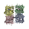
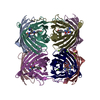
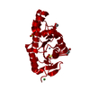
| Title | Structure and location of gene product 8 in the bacteriophage T4 baseplate | ||||||
 Components Components | baseplate structural protein gp8 | ||||||
 Keywords Keywords | VIRAL PROTEIN / bacteriophage T4 / baseplate / dimer / beta sandwich | ||||||
| Function / homology |  Function and homology information Function and homology information | ||||||
| Biological species |  Enterobacteria phage T4 (virus) Enterobacteria phage T4 (virus) | ||||||
| Method |  X-RAY DIFFRACTION / SYNCHROTRON / MAD / Resolution: 2 Å X-RAY DIFFRACTION / SYNCHROTRON / MAD / Resolution: 2 Å | ||||||
 Authors Authors | Leiman, P.G. / Shneider, M.M. / Kostyuchenko, V.A. / Chipman, P.R. / Mesyanzhinov, V.V. / Rossmann, M.G. | ||||||
 Citation Citation | Journal: J.Mol.Biol. / Year: 2003 Title: Structure and location of gene product 8 in the bacteriophage T4 baseplate Authors: Leiman, P.G. / Shneider, M.M. / Kostyuchenko, V.A. / Chipman, P.R. / Mesyanzhinov, V.V. / Rossmann, M.G. | ||||||
| History |
|
- Structure visualization
| Structure viewer | Molecule: MolmilJmol/JSmol |
|---|
- Downloads & links
-Download
| PDBx/mmCIF format | 1n7z.cif.gz | 294.4 KB | Display | PDBx/mmCIF format |
|---|---|---|---|---|
| PDB format | pdb1n7z.ent.gz | 238.2 KB | Display | PDB format |
| PDBx/mmJSON format | 1n7z.json.gz | Tree view | PDBx/mmJSON format | |
| Others |  Other downloads Other downloads |
-Validation report
| Arichive directory | https://data.pdbj.org/pub/pdb/validation_reports/n7/1n7zftp://data.pdbj.org/pub/pdb/validation_reports/n7/1n7z | HTTPS FTP |
|---|
-Related structure data


-Links
 PDBj
PDBj- Assembly
| Deposited unit | 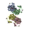
| ||||||||
|---|---|---|---|---|---|---|---|---|---|
| 1 | 
| ||||||||
| 2 | 
| ||||||||
| Unit cell |
|
-Components
| #1: Protein | Mass: 38604.406 Da / Num. of mol.: 4 Source method: isolated from a genetically manipulated source Source: (gene. exp.) Enterobacteria phage T4 (virus) / Genus: T4-like viruses / Species: Enterobacteria phage T4 sensu lato / Gene: gene 8 / Plasmid: pET-22b(+) / Production host:  #2: Chemical | ChemComp-CL /   Mass: 35.453 Da / Num. of mol.: 30 / Source method: obtained synthetically / Formula: Cl Mass: 35.453 Da / Num. of mol.: 30 / Source method: obtained synthetically / Formula: Cl#3: Water | ChemComp-HOH / |  Mass: 18.015 Da / Num. of mol.: 1025 / Source method: isolated from a natural source / Formula: H2O Mass: 18.015 Da / Num. of mol.: 1025 / Source method: isolated from a natural source / Formula: H2OHas protein modification | Y | |
|---|
-Experimental details
-Experiment
| Experiment | Method: X-RAY DIFFRACTION / Number of used crystals: 1 |
|---|
- Sample preparation
| Crystal | Density Matthews: 2.35 Å3/Da / Density % sol: 47.25 % | ||||||||||||||||||||||||||||||||||||||||||||||||||||||||
|---|---|---|---|---|---|---|---|---|---|---|---|---|---|---|---|---|---|---|---|---|---|---|---|---|---|---|---|---|---|---|---|---|---|---|---|---|---|---|---|---|---|---|---|---|---|---|---|---|---|---|---|---|---|---|---|---|---|
| Crystal grow | Temperature: 293.15 K / Method: vapor diffusion, hanging drop / pH: 7 Details: HEPES, PEG 6000, LiCl, DTT, pH 7.0, VAPOR DIFFUSION, HANGING DROP, temperature 293.15K | ||||||||||||||||||||||||||||||||||||||||||||||||||||||||
| Crystal grow | *PLUS pH: 7.5 / Method: vapor diffusion, hanging drop | ||||||||||||||||||||||||||||||||||||||||||||||||||||||||
| Components of the solutions | *PLUS
|
-Data collection
| Diffraction | Mean temperature: 100 K | ||||||||||||
|---|---|---|---|---|---|---|---|---|---|---|---|---|---|
| Diffraction source | Source: SYNCHROTRON / Site: APS  / Beamline: 14-BM-D / Wavelength: 0.9797, 0.9799,0.9426 / Beamline: 14-BM-D / Wavelength: 0.9797, 0.9799,0.9426 | ||||||||||||
| Detector | Type: ADSC QUANTUM 4 / Detector: CCD / Date: Dec 15, 2001 / Details: Bent cylindrical Si-mirror (Rh coating) | ||||||||||||
| Radiation | Monochromator: Si(111) double-crystal / Protocol: MAD / Monochromatic (M) / Laue (L): M / Scattering type: x-ray | ||||||||||||
| Radiation wavelength |
| ||||||||||||
| Reflection | Resolution: 2→50 Å / Num. all: 99719 / Num. obs: 97218 / % possible obs: 97.5 % / Observed criterion σ(F): 0 / Observed criterion σ(I): 0 / Redundancy: 1.9 % / Biso Wilson estimate: 18.1 Å2 / Rmerge(I) obs: 0.07 / Net I/σ(I): 15.1 | ||||||||||||
| Reflection shell | Resolution: 2→2.03 Å / Redundancy: 1.6 % / Rmerge(I) obs: 0.297 / Mean I/σ(I) obs: 4.2 / Num. unique all: 4777 / % possible all: 96.5 | ||||||||||||
| Reflection | *PLUS Rmerge(I) obs: 0.07 | ||||||||||||
| Reflection shell | *PLUS % possible obs: 96.5 % / Redundancy: 1.8 % |
- Processing
| Software |
| ||||||||||||||||||||||||||||||||||||||||||||||||||||||||||||||||||||||||||||||||
|---|---|---|---|---|---|---|---|---|---|---|---|---|---|---|---|---|---|---|---|---|---|---|---|---|---|---|---|---|---|---|---|---|---|---|---|---|---|---|---|---|---|---|---|---|---|---|---|---|---|---|---|---|---|---|---|---|---|---|---|---|---|---|---|---|---|---|---|---|---|---|---|---|---|---|---|---|---|---|---|---|---|
| Refinement | Method to determine structure: MAD / Resolution: 2→27.08 Å / Rfactor Rfree error: 0.005 / Data cutoff high absF: 1338852.16 / Data cutoff high rms absF: 1338852.16 / Data cutoff low absF: 0 / Isotropic thermal model: RESTRAINED / Cross valid method: THROUGHOUT / σ(F): 0 / Stereochemistry target values: Engh & Huber
| ||||||||||||||||||||||||||||||||||||||||||||||||||||||||||||||||||||||||||||||||
| Solvent computation | Solvent model: FLAT MODEL / Bsol: 33.0041 Å2 / ksol: 0.315156 e/Å3 | ||||||||||||||||||||||||||||||||||||||||||||||||||||||||||||||||||||||||||||||||
| Displacement parameters | Biso mean: 31.4 Å2
| ||||||||||||||||||||||||||||||||||||||||||||||||||||||||||||||||||||||||||||||||
| Refine analyze |
| ||||||||||||||||||||||||||||||||||||||||||||||||||||||||||||||||||||||||||||||||
| Refinement step | Cycle: LAST / Resolution: 2→27.08 Å
| ||||||||||||||||||||||||||||||||||||||||||||||||||||||||||||||||||||||||||||||||
| Refine LS restraints |
| ||||||||||||||||||||||||||||||||||||||||||||||||||||||||||||||||||||||||||||||||
| LS refinement shell | Resolution: 2→2.09 Å / Rfactor Rfree error: 0.017 / Total num. of bins used: 8
| ||||||||||||||||||||||||||||||||||||||||||||||||||||||||||||||||||||||||||||||||
| Xplor file |
| ||||||||||||||||||||||||||||||||||||||||||||||||||||||||||||||||||||||||||||||||
| Refinement | *PLUS Highest resolution: 2 Å / Lowest resolution: 40 Å / Rfactor Rwork: 0.21 | ||||||||||||||||||||||||||||||||||||||||||||||||||||||||||||||||||||||||||||||||
| Solvent computation | *PLUS | ||||||||||||||||||||||||||||||||||||||||||||||||||||||||||||||||||||||||||||||||
| Displacement parameters | *PLUS | ||||||||||||||||||||||||||||||||||||||||||||||||||||||||||||||||||||||||||||||||
| Refine LS restraints | *PLUS
|
