 Movie
Movie Controller


+ Open data

- Basic information
| Entry | Database: PDB / ID: 6j68 | ||||||
|---|---|---|---|---|---|---|---|
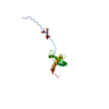
| Title | Structure of KIBRA and LATS1 Complex | ||||||
 Components Components |
| ||||||
 Keywords Keywords | CELL CYCLE / Tandem WW domain / Tandem PY motif / HIPPO Signaling | ||||||
| Function / homology |  Function and homology information Function and homology informationinner cell mass cell fate commitment / inner cell mass cellular morphogenesis / regulation of hippo signaling / Signaling by Hippo / regulation of ubiquitin-dependent protein catabolic process / regulation of transforming growth factor beta receptor signaling pathway / negative regulation of cyclin-dependent protein serine/threonine kinase activity / positive regulation of hippo signaling / sister chromatid segregation / negative regulation of organ growth ...inner cell mass cell fate commitment / inner cell mass cellular morphogenesis / regulation of hippo signaling / Signaling by Hippo / regulation of ubiquitin-dependent protein catabolic process / regulation of transforming growth factor beta receptor signaling pathway / negative regulation of cyclin-dependent protein serine/threonine kinase activity / positive regulation of hippo signaling / sister chromatid segregation / negative regulation of organ growth / NLRP3 inflammasome complex assembly / regulation of actin filament polymerization / regulation of postsynaptic density assembly / mammary gland epithelial cell differentiation / regulation of intracellular estrogen receptor signaling pathway / hippo signaling / regulation of organ growth / negative regulation of protein localization to nucleus / positive regulation of NLRP3 inflammasome complex assembly / microtubule organizing center / regulation of protein-containing complex assembly / negative regulation of hippo signaling / postsynaptic density, intracellular component / keratinocyte differentiation / hormone-mediated signaling pathway / signaling adaptor activity / nuclear estrogen receptor binding / negative regulation of canonical Wnt signaling pathway / G1/S transition of mitotic cell cycle / G2/M transition of mitotic cell cycle / ruffle membrane / kinase binding / spindle pole / intracellular protein localization / cell migration / midbody / molecular adaptor activity / protein phosphorylation / protein-macromolecule adaptor activity / positive regulation of MAPK cascade / transcription coactivator activity / non-specific serine/threonine protein kinase / postsynapse / positive regulation of apoptotic process / negative regulation of cell population proliferation / protein serine kinase activity / cell division / protein serine/threonine kinase activity / regulation of DNA-templated transcription / centrosome / protein kinase binding / perinuclear region of cytoplasm / glutamatergic synapse / magnesium ion binding / negative regulation of transcription by RNA polymerase II / protein-containing complex / ATP binding / nucleus / cytoplasm / cytosol Similarity search - Function | ||||||
| Biological species |  | ||||||
| Method |  X-RAY DIFFRACTION / SYNCHROTRON / SAD / Resolution: 2.495 Å X-RAY DIFFRACTION / SYNCHROTRON / SAD / Resolution: 2.495 Å | ||||||
 Authors Authors | Lin, Z. / Yang, Z. / Ji, Z. / Zhang, M. | ||||||
 Citation Citation | Journal: Elife / Year: 2019 Title: Decoding WW domain tandem-mediated target recognitions in tissue growth and cell polarity. Authors: Lin, Z. / Yang, Z. / Xie, R. / Ji, Z. / Guan, K. / Zhang, M. | ||||||
| History |
|
- Structure visualization
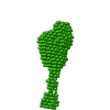
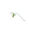
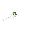
| Structure viewer | Molecule: MolmilJmol/JSmol |
|---|
- Downloads & links
-Download
| PDBx/mmCIF format | 6j68.cif.gz | 123.1 KB | Display | PDBx/mmCIF format |
|---|---|---|---|---|
| PDB format | pdb6j68.ent.gz | 95.4 KB | Display | PDB format |
| PDBx/mmJSON format | 6j68.json.gz | Tree view | PDBx/mmJSON format | |
| Others |  Other downloads Other downloads |
-Validation report
| Arichive directory | https://data.pdbj.org/pub/pdb/validation_reports/j6/6j68ftp://data.pdbj.org/pub/pdb/validation_reports/j6/6j68 | HTTPS FTP |
|---|
-Related structure data
| Related structure data | 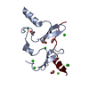 6jjwC 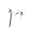 6jjxC 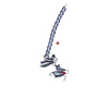 6jjyC 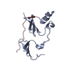 6jjzC 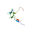 6jk0C 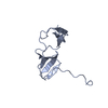 6jk1C C: citing same article ( |
|---|---|
| Similar structure data |
-Links
 PDBj
PDBj


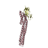
- Assembly
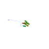
| Deposited unit | 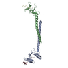
| ||||||||
|---|---|---|---|---|---|---|---|---|---|
| 1 | 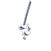
| ||||||||
| 2 | 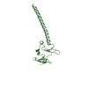
| ||||||||
| Unit cell |
|
-Components
| #1: Protein | Mass: 16036.611 Da / Num. of mol.: 2 Source method: isolated from a genetically manipulated source Source: (gene. exp.)  #2: Protein/peptide | Mass: 2926.220 Da / Num. of mol.: 2 Source method: isolated from a genetically manipulated source Source: (gene. exp.) References: UniProt: Q8BYR2, non-specific serine/threonine protein kinase #3: Water | ChemComp-HOH / |  Mass: 18.015 Da / Num. of mol.: 80 / Source method: isolated from a natural source / Formula: H2O Mass: 18.015 Da / Num. of mol.: 80 / Source method: isolated from a natural source / Formula: H2O |
|---|
-Experimental details
-Experiment
| Experiment | Method: X-RAY DIFFRACTION / Number of used crystals: 1 |
|---|
- Sample preparation
| Crystal | Density Matthews: 3.07 Å3/Da / Density % sol: 59.94 % |
|---|---|
| Crystal grow | Temperature: 289 K / Method: vapor diffusion / pH: 8 / Details: 0.1M Tris, 28% PEG 4000 |
-Data collection
| Diffraction | Mean temperature: 100 K / Serial crystal experiment: N |
|---|---|
| Diffraction source | Source: SYNCHROTRON / Site: SSRF  / Beamline: BL19U1 / Wavelength: 0.97854 Å / Beamline: BL19U1 / Wavelength: 0.97854 Å |
| Detector | Type: ADSC QUANTUM 315r / Detector: CCD / Date: Jul 13, 2015 |
| Radiation | Protocol: SINGLE WAVELENGTH / Monochromatic (M) / Laue (L): M / Scattering type: x-ray |
| Radiation wavelength | Wavelength: 0.97854 Å / Relative weight: 1 |
| Reflection | Resolution: 2.495→50 Å / Num. obs: 15953 / % possible obs: 99.5 % / Redundancy: 11.2 % / Rmerge(I) obs: 0.102 / Net I/σ(I): 23.6 |
| Reflection shell | Resolution: 2.495→2.54 Å / Rmerge(I) obs: 0.982 / Num. unique obs: 713 |
- Processing
| Software |
| ||||||||||||||||||||||||||||||||||||||||||
|---|---|---|---|---|---|---|---|---|---|---|---|---|---|---|---|---|---|---|---|---|---|---|---|---|---|---|---|---|---|---|---|---|---|---|---|---|---|---|---|---|---|---|---|
| Refinement | Method to determine structure: SAD / Resolution: 2.495→49.035 Å / SU ML: 0.37 / Cross valid method: FREE R-VALUE / σ(F): 1.34 / Phase error: 27.81 / Stereochemistry target values: ML
| ||||||||||||||||||||||||||||||||||||||||||
| Solvent computation | Shrinkage radii: 0.9 Å / VDW probe radii: 1.11 Å / Solvent model: FLAT BULK SOLVENT MODEL | ||||||||||||||||||||||||||||||||||||||||||
| Refinement step | Cycle: LAST / Resolution: 2.495→49.035 Å
| ||||||||||||||||||||||||||||||||||||||||||
| Refine LS restraints |
| ||||||||||||||||||||||||||||||||||||||||||
| LS refinement shell |
| ||||||||||||||||||||||||||||||||||||||||||
| Refinement TLS params. | Method: refined / Origin x: 13.3476 Å / Origin y: -5.0154 Å / Origin z: -20.1226 Å
| ||||||||||||||||||||||||||||||||||||||||||
| Refinement TLS group | Selection details: all |
