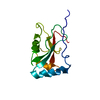
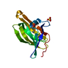
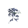
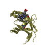
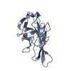
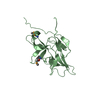
- PDB-6jf2: Crystal structure of a 20kDa fragment of FlgG -
+
Open data
ID or keywords:
Loading...
-
Basic information
Entry
Database: PDB / ID: 6jf2
Title
Crystal structure of a 20kDa fragment of FlgG
Components
Flagellar basal-body rod protein FlgG
Keywords
STRUCTURAL PROTEIN / Bacterial flagellum / distal rod
Function / homology
Function and homology information
bacterial-type flagellum basal body, distal rod / bacterial-type flagellum-dependent swarming motility / bacterial-type flagellum Similarity search - Function
Flagellar basal-body rod FlgG / Flagellar hook-basal body protein, FlgE/F/G / Flagellar hook-basal body protein, FlgE/F/G-like / : / Flagellar hook protein FlgE/F/G D1 domain / Flagellar basal body rod protein, conserved site / Flagella basal body rod proteins signature. / Flagellar basal body rod protein, N-terminal / Flagellar basal-body/hook protein, C-terminal domain / Flagella basal body rod protein / Flagellar basal body rod FlgEFG protein C-terminal Similarity search - Domain/homology
Journal: Biomolecules / Year: 2019 Title: Architecture of the Bacterial Flagellar Distal Rod and Hook of . Authors: Yumiko Saijo-Hamano / Hideyuki Matsunami / Keiichi Namba / Katsumi Imada / Abstract: The bacterial flagellum is a large molecular complex composed of thousands of protein subunits for motility. The filamentous part of the flagellum, which is called the axial structure, consists of ...The bacterial flagellum is a large molecular complex composed of thousands of protein subunits for motility. The filamentous part of the flagellum, which is called the axial structure, consists of the filament, the hook, and the rods, with other minor components-the cap protein and the hook associated proteins. They share a common basic architecture of subunit arrangement, but each part shows quite distinct mechanical properties to achieve its specific function. The distal rod and the hook are helical assemblies of a single protein, FlgG and FlgE, respectively. They show a significant sequence similarity but have distinct mechanical characteristics. The rod is a rigid, straight cylinder, whereas the hook is a curved tube with high bending flexibility. Here, we report a structural model of the rod constructed by using the crystal structure of a core fragment of FlgG with a density map obtained previously by electron cryomicroscopy. Our structural model suggests that a segment called L-stretch plays a key role in achieving the distinct mechanical properties of the rod using a structurally similar component protein to that of the hook.
In the structure databanks used in Yorodumi, some data are registered as the other names, "COVID-19 virus" and "2019-nCoV". Here are the details of the virus and the list of structure data.
Jan 31, 2019. EMDB accession codes are about to change! (news from PDBe EMDB page)
EMDB accession codes are about to change! (news from PDBe EMDB page)
The allocation of 4 digits for EMDB accession codes will soon come to an end. Whilst these codes will remain in use, new EMDB accession codes will include an additional digit and will expand incrementally as the available range of codes is exhausted. The current 4-digit format prefixed with “EMD-” (i.e. EMD-XXXX) will advance to a 5-digit format (i.e. EMD-XXXXX), and so on. It is currently estimated that the 4-digit codes will be depleted around Spring 2019, at which point the 5-digit format will come into force.
The EM Navigator/Yorodumi systems omit the EMD- prefix.
Related info.:Q: What is EMD? / ID/Accession-code notation in Yorodumi/EM Navigator
Yorodumi is a browser for structure data from EMDB, PDB, SASBDB, etc.
This page is also the successor to EM Navigator detail page, and also detail information page/front-end page for Omokage search.
The word "yorodu" (or yorozu) is an old Japanese word meaning "ten thousand". "mi" (miru) is to see.
Related info.:EMDB / PDB / SASBDB / Comparison of 3 databanks / Yorodumi Search / Aug 31, 2016. New EM Navigator & Yorodumi / Yorodumi Papers / Jmol/JSmol / Function and homology information / Changes in new EM Navigator and Yorodumi
 Movie
Movie Controller


 Open data
 Basic information
 Components
 Keywords
 Function and homology information
 Salmonella typhimurium (bacteria)
 X-RAY DIFFRACTION /
  Authors
 Citation
 Structure visualization
 Downloads & links
 Other downloads
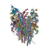


 PDBj
PDBj
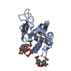
 Assembly
 Mass: 18.015 Da / Num. of mol.: 192 / Source method: isolated from a natural source / Formula: H2O
 Sample preparation
 Processing