 Movie
Movie Controller

[English] 日本語
 Yorodumi
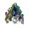
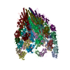
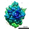
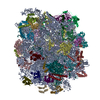
- PDB-6jzt: Structure of the bacterial flagellar hook from Salmonella typhimurium -
+ Open data

- Basic information
| Entry | Database: PDB / ID: 6jzt | ||||||||||||||||||
|---|---|---|---|---|---|---|---|---|---|---|---|---|---|---|---|---|---|---|---|
| Title | Structure of the bacterial flagellar hook from Salmonella typhimurium | ||||||||||||||||||
 Components Components | Flagellar hook protein FlgE | ||||||||||||||||||
 Keywords Keywords | MOTOR PROTEIN / Bacterial flagellum / hook protein / STRUCTURAL PROTEIN | ||||||||||||||||||
| Function / homology |  Function and homology information Function and homology informationbacterial-type flagellum hook / bacterial-type flagellum basal body / bacterial-type flagellum-dependent swarming motility / bacterial-type flagellum / cytosol Similarity search - Function | ||||||||||||||||||
| Biological species |  Salmonella typhimurium (bacteria) Salmonella typhimurium (bacteria) | ||||||||||||||||||
| Method | ELECTRON MICROSCOPY / helical reconstruction / cryo EM / Resolution: 7.1 Å | ||||||||||||||||||
 Authors Authors | Saijo-Hamano, Y. / Matsunami, H. / Namba, K. / Imada, K. | ||||||||||||||||||
| Funding support |  Japan, 5items Japan, 5items
| ||||||||||||||||||
 Citation Citation | Journal: Biomolecules / Year: 2019 Title: Architecture of the Bacterial Flagellar Distal Rod and Hook of . Authors: Yumiko Saijo-Hamano / Hideyuki Matsunami / Keiichi Namba / Katsumi Imada / Abstract: The bacterial flagellum is a large molecular complex composed of thousands of protein subunits for motility. The filamentous part of the flagellum, which is called the axial structure, consists of ...The bacterial flagellum is a large molecular complex composed of thousands of protein subunits for motility. The filamentous part of the flagellum, which is called the axial structure, consists of the filament, the hook, and the rods, with other minor components-the cap protein and the hook associated proteins. They share a common basic architecture of subunit arrangement, but each part shows quite distinct mechanical properties to achieve its specific function. The distal rod and the hook are helical assemblies of a single protein, FlgG and FlgE, respectively. They show a significant sequence similarity but have distinct mechanical characteristics. The rod is a rigid, straight cylinder, whereas the hook is a curved tube with high bending flexibility. Here, we report a structural model of the rod constructed by using the crystal structure of a core fragment of FlgG with a density map obtained previously by electron cryomicroscopy. Our structural model suggests that a segment called L-stretch plays a key role in achieving the distinct mechanical properties of the rod using a structurally similar component protein to that of the hook. | ||||||||||||||||||
| History |
|
- Structure visualization
| Movie |
Movie viewer |
|---|---|
| Structure viewer | Molecule: MolmilJmol/JSmol |
- Downloads & links
-Download
| PDBx/mmCIF format | 6jzt.cif.gz | 1.3 MB | Display | PDBx/mmCIF format |
|---|---|---|---|---|
| PDB format | pdb6jzt.ent.gz | 1.1 MB | Display | PDB format |
| PDBx/mmJSON format | 6jzt.json.gz | Tree view | PDBx/mmJSON format | |
| Others |  Other downloads Other downloads |
-Validation report
| Arichive directory | https://data.pdbj.org/pub/pdb/validation_reports/jz/6jztftp://data.pdbj.org/pub/pdb/validation_reports/jz/6jzt | HTTPS FTP |
|---|
-Related structure data
| Related structure data | 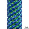 1647M  6jf2C 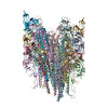 6jzrC M: map data used to model this data C: citing same article ( |
|---|---|
| Similar structure data |

-Links
 PDBj
PDBj
- Assembly
| Deposited unit | 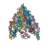
|
|---|---|
| 1 |
|
-Components
| #1: Protein | Mass: 42233.152 Da / Num. of mol.: 22 / Source method: isolated from a natural source / Source: (natural) Salmonella typhimurium (bacteria) / Variant: SJW880 / References: UniProt: A0A0J1A5C1, UniProt: P0A1J1*PLUSHas protein modification | N | |
|---|
-Experimental details
-Experiment
| Experiment | Method: ELECTRON MICROSCOPY |
|---|---|
| EM experiment | Aggregation state: FILAMENT / 3D reconstruction method: helical reconstruction |
- Sample preparation
| Component | Name: The flagellar hook / Type: COMPLEX / Entity ID: all / Source: NATURAL |
|---|---|
| Source (natural) | Organism: Salmonella enterica subsp. enterica serovar Typhimurium (bacteria) Strain: SJW880 |
| Buffer solution | pH: 8 |
| Specimen | Embedding applied: NO / Shadowing applied: NO / Staining applied: NO / Vitrification applied: YES |
| Vitrification | Cryogen name: ETHANE |
- Electron microscopy imaging
| Microscopy | Model: JEOL 3200FSC |
|---|---|
| Electron gun | Electron source:  FIELD EMISSION GUN / Accelerating voltage: 200 kV / Illumination mode: FLOOD BEAM FIELD EMISSION GUN / Accelerating voltage: 200 kV / Illumination mode: FLOOD BEAM |
| Electron lens | Mode: BRIGHT FIELD |
| Image recording | Electron dose: 20 e/Å2 / Film or detector model: TVIPS TEMCAM-F415 (4k x 4k) |
- Processing
| Software | Name: PHENIX / Version: 1.15.2_3472: / Classification: refinement | ||||||||||||||||||||||||
|---|---|---|---|---|---|---|---|---|---|---|---|---|---|---|---|---|---|---|---|---|---|---|---|---|---|
| EM software | Name: PHENIX / Category: model refinement | ||||||||||||||||||||||||
| CTF correction | Type: PHASE FLIPPING AND AMPLITUDE CORRECTION | ||||||||||||||||||||||||
| Helical symmerty | Angular rotation/subunit: 64.78 ° / Axial rise/subunit: 4.12 Å / Axial symmetry: C1 | ||||||||||||||||||||||||
| 3D reconstruction | Resolution: 7.1 Å / Resolution method: FSC 0.5 CUT-OFF / Num. of particles: 200 / Symmetry type: HELICAL | ||||||||||||||||||||||||
| Refine LS restraints |
|
