 Movie
Movie Controller


+ Open data

- Basic information
| Entry | Database: PDB / ID: 6j22 | ||||||
|---|---|---|---|---|---|---|---|
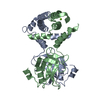
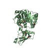
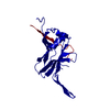
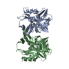
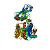
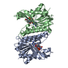
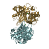
| Title | Crystal structure of Bi-functional enzyme | ||||||
 Components Components | Histidine biosynthesis bifunctional protein HisIE | ||||||
 Keywords Keywords | HYDROLASE / Bi-functional enzyme | ||||||
| Function / homology |  Function and homology information Function and homology informationphosphoribosyl-AMP cyclohydrolase / phosphoribosyl-AMP cyclohydrolase activity / phosphoribosyl-ATP diphosphatase / phosphoribosyl-ATP diphosphatase activity / L-histidine biosynthetic process / ATP binding / cytoplasm Similarity search - Function | ||||||
| Biological species |  Shigella flexneri (bacteria) Shigella flexneri (bacteria) | ||||||
| Method |  X-RAY DIFFRACTION / SYNCHROTRON / Resolution: 2.2 Å X-RAY DIFFRACTION / SYNCHROTRON / Resolution: 2.2 Å | ||||||
 Authors Authors | Zhang, H. / Shang, G. / Wang, Y. | ||||||
 Citation Citation | Journal: Biochem.Biophys.Res.Commun. / Year: 2019 Title: Structural analysis of Shigella flexneri bi-functional enzyme HisIE in histidine biosynthesis. Authors: Wang, Y. / Zhang, F. / Nie, Y. / Shang, G. / Zhang, H. | ||||||
| History |
|
- Structure visualization
| Structure viewer | Molecule: MolmilJmol/JSmol |
|---|
- Downloads & links
-Download
| PDBx/mmCIF format | 6j22.cif.gz | 91.1 KB | Display | PDBx/mmCIF format |
|---|---|---|---|---|
| PDB format | pdb6j22.ent.gz | 69.9 KB | Display | PDB format |
| PDBx/mmJSON format | 6j22.json.gz | Tree view | PDBx/mmJSON format | |
| Others |  Other downloads Other downloads |
-Validation report
| Arichive directory | https://data.pdbj.org/pub/pdb/validation_reports/j2/6j22ftp://data.pdbj.org/pub/pdb/validation_reports/j2/6j22 | HTTPS FTP |
|---|
-Related structure data


-Links
 PDBj
PDBj- Assembly
| Deposited unit | 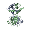
| ||||||||
|---|---|---|---|---|---|---|---|---|---|
| 1 |
| ||||||||
| Unit cell |
|
-Components
| #1: Protein | Mass: 22808.814 Da / Num. of mol.: 2 Source method: isolated from a genetically manipulated source Source: (gene. exp.) Shigella flexneri (bacteria) / Gene: hisI, hisIE, SF2088, S2209 / Production host: References: UniProt: P37793, phosphoribosyl-AMP cyclohydrolase, phosphoribosyl-ATP diphosphatase #2: Water | ChemComp-HOH / |  Mass: 18.015 Da / Num. of mol.: 198 / Source method: isolated from a natural source / Formula: H2O Mass: 18.015 Da / Num. of mol.: 198 / Source method: isolated from a natural source / Formula: H2O |
|---|
-Experimental details
-Experiment
| Experiment | Method: X-RAY DIFFRACTION / Number of used crystals: 1 |
|---|
- Sample preparation
| Crystal | Density Matthews: 2.67 Å3/Da / Density % sol: 53.95 % |
|---|---|
| Crystal grow | Temperature: 293 K / Method: vapor diffusion, hanging drop / pH: 4.6 Details: 0.1 M sodium acetate, pH 4.6, 1.8 M ammonium sulfate, 1% 1,2-butanediol |
-Data collection
| Diffraction | Mean temperature: 100 K / Serial crystal experiment: N |
|---|---|
| Diffraction source | Source: SYNCHROTRON / Site: SSRF  / Beamline: BL17U1 / Wavelength: 0.978 Å / Beamline: BL17U1 / Wavelength: 0.978 Å |
| Detector | Type: ADSC QUANTUM 315r / Detector: CCD / Date: Nov 21, 2017 |
| Radiation | Protocol: SINGLE WAVELENGTH / Monochromatic (M) / Laue (L): M / Scattering type: x-ray |
| Radiation wavelength | Wavelength: 0.978 Å / Relative weight: 1 |
| Reflection | Resolution: 2.2→50 Å / % possible obs: 93.6 % / Redundancy: 3.9 % / Net I/σ(I): 17.9 |
| Reflection shell | Resolution: 2.199→2.256 Å |
- Processing
| Software |
| ||||||||||||||||||||||||||||||||||||||||||||||||||||||||||||||||||||||||||||||||||||||||||||||||||||||||||||||||||||||||||||||||||||||||||||||||||||||||||||||||||||||||||||||||||||||
|---|---|---|---|---|---|---|---|---|---|---|---|---|---|---|---|---|---|---|---|---|---|---|---|---|---|---|---|---|---|---|---|---|---|---|---|---|---|---|---|---|---|---|---|---|---|---|---|---|---|---|---|---|---|---|---|---|---|---|---|---|---|---|---|---|---|---|---|---|---|---|---|---|---|---|---|---|---|---|---|---|---|---|---|---|---|---|---|---|---|---|---|---|---|---|---|---|---|---|---|---|---|---|---|---|---|---|---|---|---|---|---|---|---|---|---|---|---|---|---|---|---|---|---|---|---|---|---|---|---|---|---|---|---|---|---|---|---|---|---|---|---|---|---|---|---|---|---|---|---|---|---|---|---|---|---|---|---|---|---|---|---|---|---|---|---|---|---|---|---|---|---|---|---|---|---|---|---|---|---|---|---|---|---|
| Refinement | Resolution: 2.2→50 Å / Cor.coef. Fo:Fc: 0.941 / Cor.coef. Fo:Fc free: 0.899 / SU B: 6.177 / SU ML: 0.158 / Cross valid method: THROUGHOUT / ESU R: 0.267 / ESU R Free: 0.224 / Stereochemistry target values: MAXIMUM LIKELIHOOD / Details: HYDROGENS HAVE BEEN ADDED IN THE RIDING POSITIONS
| ||||||||||||||||||||||||||||||||||||||||||||||||||||||||||||||||||||||||||||||||||||||||||||||||||||||||||||||||||||||||||||||||||||||||||||||||||||||||||||||||||||||||||||||||||||||
| Solvent computation | Ion probe radii: 0.8 Å / Shrinkage radii: 0.8 Å / VDW probe radii: 1.2 Å / Solvent model: MASK | ||||||||||||||||||||||||||||||||||||||||||||||||||||||||||||||||||||||||||||||||||||||||||||||||||||||||||||||||||||||||||||||||||||||||||||||||||||||||||||||||||||||||||||||||||||||
| Displacement parameters | Biso mean: 37.516 Å2
| ||||||||||||||||||||||||||||||||||||||||||||||||||||||||||||||||||||||||||||||||||||||||||||||||||||||||||||||||||||||||||||||||||||||||||||||||||||||||||||||||||||||||||||||||||||||
| Refinement step | Cycle: 1 / Resolution: 2.2→50 Å
| ||||||||||||||||||||||||||||||||||||||||||||||||||||||||||||||||||||||||||||||||||||||||||||||||||||||||||||||||||||||||||||||||||||||||||||||||||||||||||||||||||||||||||||||||||||||
| Refine LS restraints |
|