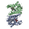
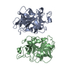
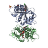
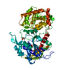
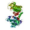
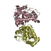
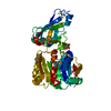
Entry Database : PDB / ID : 6l0tTitle Crystal structure of senecavirus A 3C protease 3C PROTEASE Keywords / Function / homology Function Domain/homology Component
/ / / / / / / / / / / / / / / / / / / / / / / / / / / / / / / / / / / / / / / / / / / / / / / / / / / / / / / / / / / / / / / / / / / / / / / / / Biological species Method / / / Resolution : 1.9 Å Authors Meng, K.W. / Zhang, L.J. / Meng, G. Funding support Organization Grant number Country Other government 19226631D
Journal : J.Virol. / Year : 2022Title : Structure of Senecavirus A 3C Protease Revealed the Cleavage Pattern of 3C Protease in PicornavirusesAuthors : Meng, K.W. / Zhang, L.J. / Meng, G. History Deposition Sep 27, 2019 Deposition site / Processing site Revision 1.0 Sep 30, 2020 Provider / Type Revision 2.0 Jun 15, 2022 Group Advisory / Atomic model ... Advisory / Atomic model / Author supporting evidence / Data collection / Database references / Derived calculations / Experimental preparation / Other / Polymer sequence / Refinement description / Source and taxonomy / Structure summary Category atom_site / atom_sites ... atom_site / atom_sites / cell / citation / database_2 / entity / entity_poly / entity_poly_seq / entity_src_gen / exptl_crystal / pdbx_audit_support / pdbx_contact_author / pdbx_distant_solvent_atoms / pdbx_nonpoly_scheme / pdbx_poly_seq_scheme / pdbx_struct_assembly_prop / pdbx_struct_sheet_hbond / pdbx_unobs_or_zero_occ_atoms / pdbx_unobs_or_zero_occ_residues / pdbx_validate_close_contact / pdbx_validate_rmsd_bond / pdbx_validate_symm_contact / pdbx_validate_torsion / refine / refine_hist / refine_ls_restr / refine_ls_shell / reflns / reflns_shell / software / struct_conf / struct_ref_seq / struct_ref_seq_dif / struct_sheet_range / symmetry Item _atom_sites.fract_transf_matrix[2][1] / _atom_sites.fract_transf_matrix[3][2] ... _atom_sites.fract_transf_matrix[2][1] / _atom_sites.fract_transf_matrix[3][2] / _cell.volume / _citation.country / _citation.journal_abbrev / _citation.journal_id_ASTM / _citation.journal_id_CSD / _citation.journal_id_ISSN / _citation.pdbx_database_id_DOI / _citation.title / _citation.year / _database_2.pdbx_DOI / _database_2.pdbx_database_accession / _entity.formula_weight / _entity.pdbx_description / _entity.pdbx_number_of_molecules / _entity_poly.pdbx_seq_one_letter_code / _entity_poly.pdbx_seq_one_letter_code_can / _entity_src_gen.pdbx_end_seq_num / _exptl_crystal.density_Matthews / _exptl_crystal.density_percent_sol / _pdbx_struct_assembly_prop.value / _pdbx_struct_sheet_hbond.range_1_auth_comp_id / _pdbx_struct_sheet_hbond.range_1_auth_seq_id / _pdbx_struct_sheet_hbond.range_1_label_comp_id / _pdbx_struct_sheet_hbond.range_1_label_seq_id / _pdbx_struct_sheet_hbond.range_2_auth_comp_id / _pdbx_struct_sheet_hbond.range_2_auth_seq_id / _pdbx_struct_sheet_hbond.range_2_label_comp_id / _pdbx_struct_sheet_hbond.range_2_label_seq_id / _refine.B_iso_max / _refine.B_iso_mean / _refine.B_iso_min / _refine.aniso_B[1][1] / _refine.aniso_B[1][2] / _refine.aniso_B[1][3] / _refine.aniso_B[2][2] / _refine.aniso_B[2][3] / _refine.aniso_B[3][3] / _refine.correlation_coeff_Fo_to_Fc / _refine.correlation_coeff_Fo_to_Fc_free / _refine.details / _refine.ls_R_factor_R_free / _refine.ls_R_factor_R_work / _refine.ls_R_factor_obs / _refine.ls_d_res_low / _refine.ls_number_reflns_R_free / _refine.ls_number_reflns_R_work / _refine.ls_number_reflns_obs / _refine.ls_percent_reflns_R_free / _refine.ls_percent_reflns_obs / _refine.overall_SU_B / _refine.overall_SU_ML / _refine.pdbx_ls_cross_valid_method / _refine.pdbx_ls_sigma_F / _refine.pdbx_overall_ESU_R / _refine.pdbx_overall_ESU_R_Free / _refine.pdbx_overall_phase_error / _refine.pdbx_solvent_ion_probe_radii / _refine.pdbx_solvent_shrinkage_radii / _refine.pdbx_solvent_vdw_probe_radii / _refine.pdbx_starting_model / _refine.solvent_model_details / _refine_hist.cycle_id / _refine_hist.d_res_low / _refine_hist.number_atoms_solvent / _refine_hist.number_atoms_total / _refine_hist.pdbx_B_iso_mean_solvent / _refine_hist.pdbx_number_atoms_protein / _refine_hist.pdbx_number_residues_total / _reflns.B_iso_Wilson_estimate / _reflns.d_resolution_high / _reflns.d_resolution_low / _reflns.number_obs / _reflns.pdbx_Rmerge_I_obs / _reflns.pdbx_Rsym_value / _reflns.pdbx_netI_over_sigmaI / _reflns.pdbx_redundancy / _reflns_shell.Rmerge_I_obs / _reflns_shell.d_res_high / _reflns_shell.d_res_low / _reflns_shell.number_unique_obs / _reflns_shell.pdbx_Rsym_value / _struct_conf.beg_label_seq_id / _struct_conf.end_label_seq_id / _struct_ref_seq.seq_align_beg / _struct_ref_seq.seq_align_end / _struct_sheet_range.beg_auth_comp_id / _struct_sheet_range.beg_auth_seq_id / _struct_sheet_range.beg_label_comp_id / _struct_sheet_range.beg_label_seq_id / _struct_sheet_range.end_auth_comp_id / _struct_sheet_range.end_auth_seq_id / _struct_sheet_range.end_label_comp_id / _struct_sheet_range.end_label_seq_id / _symmetry.space_group_name_Hall Description / Provider / Type Revision 2.1 Nov 22, 2023 Group / Refinement descriptionCategory / chem_comp_bond / pdbx_initial_refinement_model
Show all Show less
 Movie
Movie Controller


 Open data
 Basic information
 Components
 Keywords
 Function and homology information
 Senecavirus A
 X-RAY DIFFRACTION /
  Authors
 China, 1items
  Citation
 Structure visualization
 Downloads & links
 Other downloads
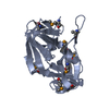


 PDBj
PDBj

 Assembly
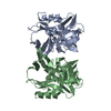

 Mass: 18.015 Da / Num. of mol.: 561 / Source method: isolated from a natural source / Formula: H2O
 Sample preparation
 Processing