 Movie
Movie Controller


+ Open data

- Basic information
| Entry | Database: PDB / ID: 3ip2 | |||||||||
|---|---|---|---|---|---|---|---|---|---|---|
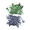
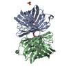
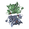
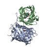
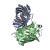
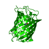
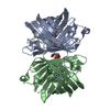
| Title | Crystal structure of red fluorescent protein Neptune at pH 7.0 | |||||||||
 Components Components | Neptune red fluorescent protein | |||||||||
 Keywords Keywords | FLUORESCENT PROTEIN | |||||||||
| Function / homology | Green Fluorescent Protein / Green fluorescent protein / Green fluorescent protein-related / Green fluorescent protein / Green fluorescent protein / bioluminescence / Beta Barrel / Mainly Beta / Red fluorescent protein eqFP611 Function and homology information Function and homology information | |||||||||
| Biological species |  Entacmaea quadricolor (sea anemone) Entacmaea quadricolor (sea anemone) | |||||||||
| Method |  X-RAY DIFFRACTION / SYNCHROTRON / MOLECULAR REPLACEMENT / Resolution: 1.6 Å X-RAY DIFFRACTION / SYNCHROTRON / MOLECULAR REPLACEMENT / Resolution: 1.6 Å | |||||||||
 Authors Authors | Lin, M.Z. / McKeown, M.R. / Ng, H.L. / Aguilera, T.A. / Shaner, N.C. / Ma, W. / Adams, S.R. / Campbell, R.E. / Alber, T. / Tsien, R.Y. | |||||||||
 Citation Citation | Journal: Chem.Biol. / Year: 2009 Title: Autofluorescent proteins with excitation in the optical window for intravital imaging in mammals. Authors: Lin, M.Z. / McKeown, M.R. / Ng, H.L. / Aguilera, T.A. / Shaner, N.C. / Campbell, R.E. / Adams, S.R. / Gross, L.A. / Ma, W. / Alber, T. / Tsien, R.Y. | |||||||||
| History |
|
- Structure visualization
| Structure viewer | Molecule: MolmilJmol/JSmol |
|---|
- Downloads & links
-Download
| PDBx/mmCIF format | 3ip2.cif.gz | 116.1 KB | Display | PDBx/mmCIF format |
|---|---|---|---|---|
| PDB format | pdb3ip2.ent.gz | 90 KB | Display | PDB format |
| PDBx/mmJSON format | 3ip2.json.gz | Tree view | PDBx/mmJSON format | |
| Others |  Other downloads Other downloads |
-Validation report
| Arichive directory | https://data.pdbj.org/pub/pdb/validation_reports/ip/3ip2ftp://data.pdbj.org/pub/pdb/validation_reports/ip/3ip2 | HTTPS FTP |
|---|
-Related structure data
| Related structure data |  3bxaS S: Starting model for refinement |
|---|---|
| Similar structure data |


-Links
 PDBj
PDBj

- Assembly
| Deposited unit | 
| ||||||||
|---|---|---|---|---|---|---|---|---|---|
| 1 | 
| ||||||||
| Unit cell |
| ||||||||
| Components on special symmetry positions |
| ||||||||
| Details | authors state that this is a WEAK DIMER (DISSOCIATION CONSTANT 500 NANOMOLAR). |
-Components
| #1: Protein | Mass: 27369.211 Da / Num. of mol.: 1 Source method: isolated from a genetically manipulated source Source: (gene. exp.) Entacmaea quadricolor (sea anemone) / Production host:  |
|---|---|
| #2: Water | ChemComp-HOH /  Mass: 18.015 Da / Num. of mol.: 212 / Source method: isolated from a natural source / Formula: H2O Mass: 18.015 Da / Num. of mol.: 212 / Source method: isolated from a natural source / Formula: H2O |
| Has protein modification | Y |
| Sequence details | RESIDUES MET 63, TYR 64 AND GLY 65 AUTOCATALYTICALLY FORM THE CHROMOPHORE NRQ LABELLED AS RESIDUE ...RESIDUES MET 63, TYR 64 AND GLY 65 AUTOCATALY |
-Experimental details
-Experiment
| Experiment | Method: X-RAY DIFFRACTION / Number of used crystals: 1 |
|---|
- Sample preparation
| Crystal | Density Matthews: 2.06 Å3/Da / Density % sol: 40.39 % |
|---|---|
| Crystal grow | Temperature: 291 K / Method: vapor diffusion, sitting drop / pH: 7 Details: 1 uL of protein at 10 mg/mL was mixed with 1 uL of 0.1 M HEPES, 0.2 M CaCl2, 20% PEG 6000, pH 7.0, VAPOR DIFFUSION, SITTING DROP, temperature 291K |
-Data collection
| Diffraction | Mean temperature: 100 K |
|---|---|
| Diffraction source | Source: SYNCHROTRON / Site: ALS  / Beamline: 8.3.1 / Wavelength: 1.11586 Å / Beamline: 8.3.1 / Wavelength: 1.11586 Å |
| Detector | Type: ADSC QUANTUM 315r / Detector: CCD / Date: Apr 1, 2009 |
| Radiation | Monochromator: KHOZU Double flat crystal / Protocol: SINGLE WAVELENGTH / Monochromatic (M) / Laue (L): M / Scattering type: x-ray |
| Radiation wavelength | Wavelength: 1.11586 Å / Relative weight: 1 |
| Reflection | Resolution: 1.6→92.1 Å / Num. all: 30810 / Num. obs: 30755 / % possible obs: 99.9 % / Observed criterion σ(F): -3 / Observed criterion σ(I): -3 / Redundancy: 8.5 % / Rsym value: 0.055 / Net I/σ(I): 23.8 |
| Reflection shell | Resolution: 1.6→1.68 Å / Mean I/σ(I) obs: 2.2 / Num. unique all: 4369 / Rsym value: 0.778 / % possible all: 99.7 |
- Processing
| Software |
| ||||||||||||||||||||||||||||||||||||||||||||||||||||||||||||||||||||||||||||||||||||
|---|---|---|---|---|---|---|---|---|---|---|---|---|---|---|---|---|---|---|---|---|---|---|---|---|---|---|---|---|---|---|---|---|---|---|---|---|---|---|---|---|---|---|---|---|---|---|---|---|---|---|---|---|---|---|---|---|---|---|---|---|---|---|---|---|---|---|---|---|---|---|---|---|---|---|---|---|---|---|---|---|---|---|---|---|---|
| Refinement | Method to determine structure: MOLECULAR REPLACEMENT Starting model: pdb entry 3BXA Resolution: 1.6→65.2 Å / SU ML: 0.27 / σ(F): -3 / Stereochemistry target values: Engh & Huber / Details: TLS refinement
| ||||||||||||||||||||||||||||||||||||||||||||||||||||||||||||||||||||||||||||||||||||
| Solvent computation | Shrinkage radii: 0.9 Å / VDW probe radii: 1.11 Å / Solvent model: FLAT BULK SOLVENT MODEL / Bsol: 60.022 Å2 / ksol: 0.42 e/Å3 | ||||||||||||||||||||||||||||||||||||||||||||||||||||||||||||||||||||||||||||||||||||
| Refinement step | Cycle: LAST / Resolution: 1.6→65.2 Å
| ||||||||||||||||||||||||||||||||||||||||||||||||||||||||||||||||||||||||||||||||||||
| LS refinement shell |
| ||||||||||||||||||||||||||||||||||||||||||||||||||||||||||||||||||||||||||||||||||||
| Refinement TLS params. | Method: refined / Origin x: -0.8391 Å / Origin y: 21.7715 Å / Origin z: 25.2752 Å
| ||||||||||||||||||||||||||||||||||||||||||||||||||||||||||||||||||||||||||||||||||||
| Refinement TLS group | Selection details: chain A |
