 Movie
Movie Controller

[English] 日本語
 Yorodumi
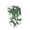
- PDB-3c8h: Crystal structure of the enterobactin esterase FES from Shigella ... -
+ Open data

- Basic information
| Entry | Database: PDB / ID: 3c8h | ||||||
|---|---|---|---|---|---|---|---|
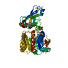
| Title | Crystal structure of the enterobactin esterase FES from Shigella flexneri in the presence of 2,3-Di-hydroxy-N-benzoyl-serine | ||||||
 Components Components | Enterochelin esterase | ||||||
 Keywords Keywords | HYDROLASE / alpha-beta-alpha sandwich / IroD / Iron aquisition / Structural Genomics / PSI-2 / Protein Structure Initiative / Midwest Center for Structural Genomics / MCSG | ||||||
| Function / homology |  Function and homology information Function and homology informationenterochelin esterase activity / iron(III)-enterobactin esterase / iron ion transport / iron ion binding / cytoplasm Similarity search - Function | ||||||
| Biological species |  Shigella flexneri 2a str. 2457T (bacteria) Shigella flexneri 2a str. 2457T (bacteria) | ||||||
| Method |  X-RAY DIFFRACTION / SYNCHROTRON / MOLECULAR REPLACEMENT / Resolution: 2.48 Å X-RAY DIFFRACTION / SYNCHROTRON / MOLECULAR REPLACEMENT / Resolution: 2.48 Å | ||||||
 Authors Authors | Kim, Y. / Maltseva, N. / Abergel, R. / Holzle, D. / Raymond, K. / Joachimiak, A. / Midwest Center for Structural Genomics (MCSG) | ||||||
 Citation Citation | Journal: To be Published Title: Siderophore Mediated Iron Acquisition: Structure and Specificity of Enterobactin Esterase from Shigella flexneri. Authors: Kim, Y. / Maltseva, N. / Abergel, R. / Holzle, D. / Raymond, K. / Joachimiak, A. | ||||||
| History |
|
- Structure visualization
| Structure viewer | Molecule: MolmilJmol/JSmol |
|---|
- Downloads & links
-Download
| PDBx/mmCIF format | 3c8h.cif.gz | 616.9 KB | Display | PDBx/mmCIF format |
|---|---|---|---|---|
| PDB format | pdb3c8h.ent.gz | 514.3 KB | Display | PDB format |
| PDBx/mmJSON format | 3c8h.json.gz | Tree view | PDBx/mmJSON format | |
| Others |  Other downloads Other downloads |
-Validation report
| Arichive directory | https://data.pdbj.org/pub/pdb/validation_reports/c8/3c8hftp://data.pdbj.org/pub/pdb/validation_reports/c8/3c8h | HTTPS FTP |
|---|
-Related structure data
| Related structure data | 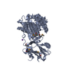 3c87C  3c8dC  2b20S S: Starting model for refinement C: citing same article ( |
|---|---|
| Similar structure data | |
| Other databases |


-Links
 PDBj
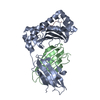
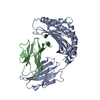
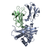
PDBj- Assembly
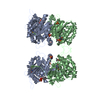
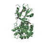
| Deposited unit | 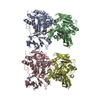
| ||||||||
|---|---|---|---|---|---|---|---|---|---|
| 1 | 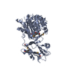
| ||||||||
| 2 | 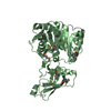
| ||||||||
| 3 | 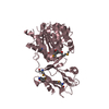
| ||||||||
| 4 | 
| ||||||||
| 5 | 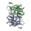
| ||||||||
| 6 | 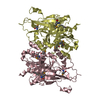
| ||||||||
| Unit cell |
|
-Components
| #1: Protein | Mass: 46304.078 Da / Num. of mol.: 4 Source method: isolated from a genetically manipulated source Source: (gene. exp.) Shigella flexneri 2a str. 2457T (bacteria)Species: Shigella flexneri / Strain: 2457T / Serotype 2a / Gene: fes, S0503, SF0497 / Plasmid: pMCSG7 / Species (production host): Escherichia coli / Production host: #2: Water | ChemComp-HOH / |  Mass: 18.015 Da / Num. of mol.: 333 / Source method: isolated from a natural source / Formula: H2O Mass: 18.015 Da / Num. of mol.: 333 / Source method: isolated from a natural source / Formula: H2OHas protein modification | Y | |
|---|
-Experimental details
-Experiment
| Experiment | Method: X-RAY DIFFRACTION / Number of used crystals: 1 |
|---|
- Sample preparation
| Crystal | Density Matthews: 2.3 Å3/Da / Density % sol: 46.44 % |
|---|---|
| Crystal grow | Temperature: 291 K / Method: vapor diffusion, sitting drop / pH: 5.6 Details: 20 % PEG 3000, 0.1 M Tri-sodium citrate pH 5.6, 3 mM DHBS, 1 mM FeCl3, VAPOR DIFFUSION, SITTING DROP, temperature 291K |
-Data collection
| Diffraction | Mean temperature: 100 K |
|---|---|
| Diffraction source | Source: SYNCHROTRON / Site: APS  / Beamline: 19-ID / Wavelength: 0.9793 Å / Beamline: 19-ID / Wavelength: 0.9793 Å |
| Detector | Type: ADSC QUANTUM 315 / Detector: CCD / Date: Nov 3, 2006 / Details: Mirrors |
| Radiation | Monochromator: Double crystal / Protocol: SINGLE WAVELENGTH / Monochromatic (M) / Laue (L): M / Scattering type: x-ray |
| Radiation wavelength | Wavelength: 0.9793 Å / Relative weight: 1 |
| Reflection | Resolution: 2.48→47.23 Å / Num. all: 57160 / Num. obs: 57160 / % possible obs: 95.2 % / Observed criterion σ(F): 0 / Observed criterion σ(I): 0 / Redundancy: 3.4 % / Biso Wilson estimate: 40.47 Å2 / Rsym value: 0.106 / Net I/σ(I): 8.6 |
| Reflection shell | Resolution: 2.48→2.59 Å / Redundancy: 2.6 % / Mean I/σ(I) obs: 2.12 / Num. unique all: 4512 / Rsym value: 0.462 / % possible all: 76 |
- Processing
| Software |
| ||||||||||||||||||||||||||||||||||||||||||||||||||||||||||||||||||||||||||||||||||||||||||||||||||||||||||||||||||||||||||||||||||
|---|---|---|---|---|---|---|---|---|---|---|---|---|---|---|---|---|---|---|---|---|---|---|---|---|---|---|---|---|---|---|---|---|---|---|---|---|---|---|---|---|---|---|---|---|---|---|---|---|---|---|---|---|---|---|---|---|---|---|---|---|---|---|---|---|---|---|---|---|---|---|---|---|---|---|---|---|---|---|---|---|---|---|---|---|---|---|---|---|---|---|---|---|---|---|---|---|---|---|---|---|---|---|---|---|---|---|---|---|---|---|---|---|---|---|---|---|---|---|---|---|---|---|---|---|---|---|---|---|---|---|---|
| Refinement | Method to determine structure: MOLECULAR REPLACEMENT Starting model: PDB entry 2B20 Resolution: 2.48→47.23 Å / Cross valid method: THROUGHOUT / σ(F): 0 / σ(I): 0 / Stereochemistry target values: Engh & Huber Details: 1. TWINNING: TWIN LAW=-H,-K,L, TWIN FRACTION: 0.502. 2. Structure has been refined using PHENIX.REFINE, PHENIX.XTRIAGE programs. 3. When refining TLS, the output PDB file always has the ...Details: 1. TWINNING: TWIN LAW=-H,-K,L, TWIN FRACTION: 0.502. 2. Structure has been refined using PHENIX.REFINE, PHENIX.XTRIAGE programs. 3. When refining TLS, the output PDB file always has the ANISOU records for the atoms involved in TLS groups. The anisotropic B-factor in ANISOU records is the total B-factor (B_tls + B_individual). The isotropic equivalent B-factor in ATOM records is the mean of the trace of the ANISOU matrix divided by 10000 and multiplied by 8*pi^2 and represents the isotropic equivalent of the total B-factor (B_tls + B_individual). To obtain the individual B-factors, one needs to compute the TLS component (B_tls) using the TLS records in the PDB file header and then subtract it from the total B-factors (on the ANISOU records).
| ||||||||||||||||||||||||||||||||||||||||||||||||||||||||||||||||||||||||||||||||||||||||||||||||||||||||||||||||||||||||||||||||||
| Solvent computation | Bsol: 53.56 Å2 / ksol: 0.36 e/Å3 | ||||||||||||||||||||||||||||||||||||||||||||||||||||||||||||||||||||||||||||||||||||||||||||||||||||||||||||||||||||||||||||||||||
| Displacement parameters | Biso mean: 56.15 Å2
| ||||||||||||||||||||||||||||||||||||||||||||||||||||||||||||||||||||||||||||||||||||||||||||||||||||||||||||||||||||||||||||||||||
| Refinement step | Cycle: LAST / Resolution: 2.48→47.23 Å
| ||||||||||||||||||||||||||||||||||||||||||||||||||||||||||||||||||||||||||||||||||||||||||||||||||||||||||||||||||||||||||||||||||
| Refine LS restraints |
| ||||||||||||||||||||||||||||||||||||||||||||||||||||||||||||||||||||||||||||||||||||||||||||||||||||||||||||||||||||||||||||||||||
| LS refinement shell | Refine-ID: X-RAY DIFFRACTION / Rfactor Rfree: 0.32 / Total num. of bins used: 20
| ||||||||||||||||||||||||||||||||||||||||||||||||||||||||||||||||||||||||||||||||||||||||||||||||||||||||||||||||||||||||||||||||||
| Refinement TLS params. | Refine-ID: X-RAY DIFFRACTION
| ||||||||||||||||||||||||||||||||||||||||||||||||||||||||||||||||||||||||||||||||||||||||||||||||||||||||||||||||||||||||||||||||||
| Refinement TLS group |
|
