 Movie
Movie Controller


+ Open data

- Basic information
| Entry | Database: PDB / ID: 4e0r | ||||||
|---|---|---|---|---|---|---|---|
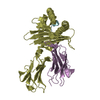
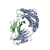
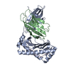
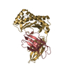
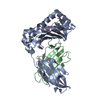
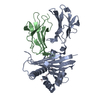
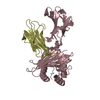
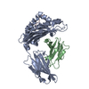
| Title | Structure of the chicken MHC class I molecule BF2*0401 | ||||||
 Components Components |
| ||||||
 Keywords Keywords | IMMUNE SYSTEM / MHC I COMPLEX / NARROW BINDING GROOVE | ||||||
| Function / homology |  Function and homology information Function and homology informationMHC class I protein binding, via antigen binding groove / ER-Phagosome pathway / Endosomal/Vacuolar pathway / Immunoregulatory interactions between a Lymphoid and a non-Lymphoid cell / DAP12 signaling / Antigen Presentation: Folding, assembly and peptide loading of class I MHC / antigen processing and presentation of peptide antigen via MHC class I / RNA processing / Neutrophil degranulation / negative regulation of iron ion transport ...MHC class I protein binding, via antigen binding groove / ER-Phagosome pathway / Endosomal/Vacuolar pathway / Immunoregulatory interactions between a Lymphoid and a non-Lymphoid cell / DAP12 signaling / Antigen Presentation: Folding, assembly and peptide loading of class I MHC / antigen processing and presentation of peptide antigen via MHC class I / RNA processing / Neutrophil degranulation / negative regulation of iron ion transport / negative regulation of forebrain neuron differentiation / peptide antigen assembly with MHC class I protein complex / HFE-transferrin receptor complex / MHC class I peptide loading complex / transferrin transport / cellular response to iron ion / negative regulation of receptor-mediated endocytosis / positive regulation of T cell cytokine production / antigen processing and presentation of endogenous peptide antigen via MHC class I / MHC class I protein complex / peptide antigen assembly with MHC class II protein complex / negative regulation of neurogenesis / MHC class II protein complex / cellular response to nicotine / positive regulation of receptor-mediated endocytosis / antigen processing and presentation of exogenous peptide antigen via MHC class II / positive regulation of immune response / peptide antigen binding / positive regulation of T cell activation / negative regulation of epithelial cell proliferation / positive regulation of cellular senescence / MHC class II protein complex binding / late endosome membrane / amyloid fibril formation / protein homotetramerization / intracellular iron ion homeostasis / learning or memory / immune response / lysosomal membrane / regulation of DNA-templated transcription / structural molecule activity / Golgi apparatus / protein homodimerization activity / DNA binding / RNA binding / extracellular region / zinc ion binding / nucleus / cytosol Similarity search - Function | ||||||
| Biological species |  | ||||||
| Method |  X-RAY DIFFRACTION / MOLECULAR REPLACEMENT / Resolution: 2.26 Å X-RAY DIFFRACTION / MOLECULAR REPLACEMENT / Resolution: 2.26 Å | ||||||
 Authors Authors | Zhang, J. / Chen, Y. / Qi, J. / Gao, F. / Kaufman, J. / Xia, C. / Gao, G.F. | ||||||
 Citation Citation | Journal: J.Immunol. / Year: 2012 Title: Narrow Groove and Restricted Anchors of MHC Class I Molecule BF2*0401 Plus Peptide Transporter Restriction Can Explain Disease Susceptibility of B4 Chickens. Authors: Zhang, J. / Chen, Y. / Qi, J. / Gao, F. / Liu, Y. / Liu, J. / Zhou, X. / Kaufman, J. / Xia, C. / Gao, G.F. | ||||||
| History |
|
- Structure visualization
| Structure viewer | Molecule: MolmilJmol/JSmol |
|---|
- Downloads & links
-Download
| PDBx/mmCIF format | 4e0r.cif.gz | 173.1 KB | Display | PDBx/mmCIF format |
|---|---|---|---|---|
| PDB format | pdb4e0r.ent.gz | 137.4 KB | Display | PDB format |
| PDBx/mmJSON format | 4e0r.json.gz | Tree view | PDBx/mmJSON format | |
| Others |  Other downloads Other downloads |
-Validation report
| Arichive directory | https://data.pdbj.org/pub/pdb/validation_reports/e0/4e0rftp://data.pdbj.org/pub/pdb/validation_reports/e0/4e0r | HTTPS FTP |
|---|
-Related structure data
| Related structure data | 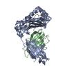 4g42C 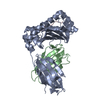 4g43C 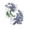 3bevS S: Starting model for refinement C: citing same article ( |
|---|---|
| Similar structure data |


-Links
 PDBj
PDBj

- Assembly
| Deposited unit | 
| ||||||||
|---|---|---|---|---|---|---|---|---|---|
| 1 | 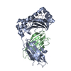
| ||||||||
| 2 | 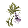
| ||||||||
| Unit cell |
|
-Components
| #1: Protein | Mass: 31853.465 Da / Num. of mol.: 2 / Fragment: UNP residues 22-291 / Mutation: D244E Source method: isolated from a genetically manipulated source Source: (gene. exp.)  #2: Protein | Mass: 11469.888 Da / Num. of mol.: 2 Source method: isolated from a genetically manipulated source Source: (gene. exp.) #3: Protein/peptide | Mass: 1010.078 Da / Num. of mol.: 2 / Fragment: UNP residues 319-326 / Source method: obtained synthetically / Details: chemical synthesis from Fus proto-onc gene / Source: (synth.) #4: Water | ChemComp-HOH / |  Mass: 18.015 Da / Num. of mol.: 517 / Source method: isolated from a natural source / Formula: H2O Mass: 18.015 Da / Num. of mol.: 517 / Source method: isolated from a natural source / Formula: H2OHas protein modification | Y | |
|---|
-Experimental details
-Experiment
| Experiment | Method: X-RAY DIFFRACTION / Number of used crystals: 1 |
|---|
- Sample preparation
| Crystal | Density Matthews: 2.16 Å3/Da / Density % sol: 42.99 % |
|---|---|
| Crystal grow | Temperature: 291 K / Method: vapor diffusion, hanging drop / pH: 7 Details: 0.1M HEPES BUFFER (PH 7.0), 5% MPD, 20% PEG 6000, VAPOR DIFFUSION, HANGING DROP, temperature 291K |
-Data collection
| Diffraction | Mean temperature: 100 K |
|---|---|
| Diffraction source | Source: ROTATING ANODE / Type: RIGAKU MICROMAX-007 / Wavelength: 1.5418 / Wavelength: 1.5418 Å |
| Detector | Type: RIGAKU RAXIS VII / Detector: IMAGE PLATE / Date: Jul 6, 2008 |
| Radiation | Monochromator: SI 111 CHANNEL / Protocol: SINGLE WAVELENGTH / Monochromatic (M) / Laue (L): M / Scattering type: x-ray |
| Radiation wavelength | Wavelength: 1.5418 Å / Relative weight: 1 |
| Reflection | Resolution: 2.259→50 Å / Num. all: 36222 / Num. obs: 34950 / % possible obs: 96.6 % / Observed criterion σ(F): 0 / Observed criterion σ(I): -3 / Redundancy: 3.1 % / Rmerge(I) obs: 0.055 / Rsym value: 0.055 / Net I/σ(I): 19.6 |
| Reflection shell | Resolution: 2.26→2.36 Å / Redundancy: 3.1 % / Rmerge(I) obs: 0.196 / Mean I/σ(I) obs: 6.8 / Rsym value: 0.196 / % possible all: 94 |
- Processing
| Software |
| ||||||||||||||||||||||||||||||||||||||||||||||||||||||||||||||||||||||||||||||
|---|---|---|---|---|---|---|---|---|---|---|---|---|---|---|---|---|---|---|---|---|---|---|---|---|---|---|---|---|---|---|---|---|---|---|---|---|---|---|---|---|---|---|---|---|---|---|---|---|---|---|---|---|---|---|---|---|---|---|---|---|---|---|---|---|---|---|---|---|---|---|---|---|---|---|---|---|---|---|---|
| Refinement | Method to determine structure: MOLECULAR REPLACEMENT Starting model: PDB ENTRY 3BEV Resolution: 2.26→43.83 Å / SU ML: 0.34 / σ(F): 1.34 / Phase error: 26.83 / Stereochemistry target values: ML / Details: REFMAC WAS ALSO USED IN REFINEMENT.
| ||||||||||||||||||||||||||||||||||||||||||||||||||||||||||||||||||||||||||||||
| Solvent computation | Shrinkage radii: 0.9 Å / VDW probe radii: 1.11 Å / Solvent model: FLAT BULK SOLVENT MODEL / Bsol: 39.95 Å2 / ksol: 0.35 e/Å3 | ||||||||||||||||||||||||||||||||||||||||||||||||||||||||||||||||||||||||||||||
| Displacement parameters | Biso mean: 28.05 Å2
| ||||||||||||||||||||||||||||||||||||||||||||||||||||||||||||||||||||||||||||||
| Refinement step | Cycle: LAST / Resolution: 2.26→43.83 Å
| ||||||||||||||||||||||||||||||||||||||||||||||||||||||||||||||||||||||||||||||
| Refine LS restraints |
| ||||||||||||||||||||||||||||||||||||||||||||||||||||||||||||||||||||||||||||||
| LS refinement shell | Refine-ID: X-RAY DIFFRACTION / Total num. of bins used: 12
|
