 Movie
Movie Controller

[English] 日本語
 Yorodumi
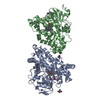
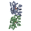
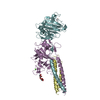
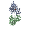
- PDB-6izv: Averaged strand structure of a 15-stranded ParM filament from Clo... -
+ Open data

- Basic information
| Entry | Database: PDB / ID: 6izv | ||||||||||||
|---|---|---|---|---|---|---|---|---|---|---|---|---|---|
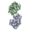
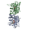
| Title | Averaged strand structure of a 15-stranded ParM filament from Clostridium botulinum | ||||||||||||
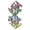
 Components Components | Putative plasmid segregation protein ParM | ||||||||||||
 Keywords Keywords | PROTEIN FIBRIL / ParM / filaments / cytoskeleton | ||||||||||||
| Function / homology | ParM-like / Actin-like protein, N-terminal / Actin like proteins N terminal domain / ATPase, nucleotide binding domain / ADENOSINE-5'-DIPHOSPHATE / Actin-like protein N-terminal domain-containing protein Function and homology information Function and homology information | ||||||||||||
| Biological species |  Clostridium botulinum Prevot_594 (bacteria) Clostridium botulinum Prevot_594 (bacteria) | ||||||||||||
| Method | ELECTRON MICROSCOPY / helical reconstruction / cryo EM / Resolution: 4.2 Å | ||||||||||||
 Authors Authors | Koh, F. / Narita, A. / Lee, L.J. / Tan, Y.Z. / Dandey, V.P. / Tanaka, K. / Popp, D. / Robinson, R.C. | ||||||||||||
| Funding support |  United States, 3items United States, 3items
| ||||||||||||
 Citation Citation | Journal: Nat Commun / Year: 2019 Title: The structure of a 15-stranded actin-like filament from Clostridium botulinum. Authors: Fujiet Koh / Akihiro Narita / Lin Jie Lee / Kotaro Tanaka / Yong Zi Tan / Venkata P Dandey / David Popp / Robert C Robinson /    Abstract: Microfilaments (actin) and microtubules represent the extremes in eukaryotic cytoskeleton cross-sectional dimensions, raising the question of whether filament architectures are limited by protein ...Microfilaments (actin) and microtubules represent the extremes in eukaryotic cytoskeleton cross-sectional dimensions, raising the question of whether filament architectures are limited by protein fold. Here, we report the cryoelectron microscopy structure of a complex filament formed from 15 protofilaments of an actin-like protein. This actin-like ParM is encoded on the large pCBH Clostridium botulinum plasmid. In cross-section, the ~26 nm diameter filament comprises a central helical protofilament surrounded by intermediate and outer layers of six and eight twisted protofilaments, respectively. Alternating polarity of the layers allows for similar lateral contacts between each layer. This filament design is stiffer than the actin filament, and has likely been selected for during evolution to move large cargos. The comparable sizes of microtubule and pCBH ParM filaments indicate that larger filament architectures are not limited by the protomer fold. Instead, function appears to have been the evolutionary driving force to produce broad, complex filaments. | ||||||||||||
| History |
|
- Structure visualization
| Movie |
Movie viewer |
|---|---|
| Structure viewer | Molecule: MolmilJmol/JSmol |
- Downloads & links
-Download
| PDBx/mmCIF format | 6izv.cif.gz | 132.6 KB | Display | PDBx/mmCIF format |
|---|---|---|---|---|
| PDB format | pdb6izv.ent.gz | 104.3 KB | Display | PDB format |
| PDBx/mmJSON format | 6izv.json.gz | Tree view | PDBx/mmJSON format | |
| Others |  Other downloads Other downloads |
-Validation report
| Arichive directory | https://data.pdbj.org/pub/pdb/validation_reports/iz/6izvftp://data.pdbj.org/pub/pdb/validation_reports/iz/6izv | HTTPS FTP |
|---|
-Related structure data
| Related structure data | 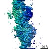 9758MC 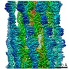 9757C  6ixwC  6izrC C: citing same article ( M: map data used to model this data |
|---|---|
| Similar structure data |


-Links
 PDBj
PDBj- Assembly
| Deposited unit | 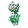
|
|---|---|
| 1 |
|
-Components
| #1: Protein | Mass: 39560.168 Da / Num. of mol.: 2 Source method: isolated from a genetically manipulated source Source: (gene. exp.) Clostridium botulinum Prevot_594 (bacteria)Gene: T258_3831 / Production host: #2: Chemical |   Mass: 427.201 Da / Num. of mol.: 2 / Source method: obtained synthetically / Formula: C10H15N5O10P2 / Comment: ADP, energy-carrying molecule*YM Mass: 427.201 Da / Num. of mol.: 2 / Source method: obtained synthetically / Formula: C10H15N5O10P2 / Comment: ADP, energy-carrying molecule*YM#3: Chemical |   Mass: 24.305 Da / Num. of mol.: 2 / Source method: obtained synthetically / Formula: Mg Mass: 24.305 Da / Num. of mol.: 2 / Source method: obtained synthetically / Formula: Mg |
|---|
-Experimental details
-Experiment
| Experiment | Method: ELECTRON MICROSCOPY |
|---|---|
| EM experiment | Aggregation state: FILAMENT / 3D reconstruction method: helical reconstruction |
- Sample preparation
| Component | Name: ParM filament coded on pCBH plasmid from Clostridium botulinum Type: COMPLEX / Entity ID: #1 / Source: RECOMBINANT |
|---|---|
| Molecular weight | Experimental value: NO |
| Source (natural) | Organism: Clostridium botulinum F str. 230613 (bacteria) / Organelle: pCBH plasmid |
| Source (recombinant) | Organism: |
| Buffer solution | pH: 7.4 |
| Specimen | Conc.: 0.8 mg/ml / Embedding applied: NO / Shadowing applied: NO / Staining applied: NO / Vitrification applied: YES |
| Vitrification | Cryogen name: ETHANE |
- Electron microscopy imaging
| Experimental equipment |  Model: Titan Krios / Image courtesy: FEI Company |
|---|---|
| Microscopy | Model: FEI TITAN KRIOS |
| Electron gun | Electron source:  FIELD EMISSION GUN / Accelerating voltage: 300 kV / Illumination mode: FLOOD BEAM FIELD EMISSION GUN / Accelerating voltage: 300 kV / Illumination mode: FLOOD BEAM |
| Electron lens | Mode: BRIGHT FIELD |
| Image recording | Electron dose: 30 e/Å2 / Detector mode: COUNTING / Film or detector model: GATAN K2 SUMMIT (4k x 4k) |
- Processing
| EM software | Name: RELION / Version: 2.1 / Category: 3D reconstruction |
|---|---|
| CTF correction | Type: PHASE FLIPPING AND AMPLITUDE CORRECTION |
| Helical symmerty | Angular rotation/subunit: -50.37 ° / Axial rise/subunit: 50.26 Å / Axial symmetry: C1 |
| 3D reconstruction | Resolution: 4.2 Å / Resolution method: OTHER / Num. of particles: 33356 / Symmetry type: HELICAL |
