 Movie
Movie Controller

[English] 日本語
 Yorodumi
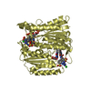
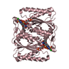
- PDB-6jxn: Crystal Structure of Indigo reductase from Bacillus smithii type ... -
+ Open data

- Basic information
| Entry | Database: PDB / ID: 6jxn | ||||||
|---|---|---|---|---|---|---|---|
| Title | Crystal Structure of Indigo reductase from Bacillus smithii type strain DSM 4216 | ||||||
 Components Components | FMN-dependent NADH-azoreductase | ||||||
 Keywords Keywords | OXIDOREDUCTASE / Indigo reductase Aizome (indigo dyeing) | ||||||
| Function / homology |  Function and homology information Function and homology informationFMN-dependent NADH-azoreductase / oxidoreductase activity, acting on NAD(P)H as acceptor / Oxidoreductases; Acting on NADH or NADPH; With a quinone or similar compound as acceptor / oxidoreductase activity, acting on NAD(P)H, quinone or similar compound as acceptor / FMN binding / electron transfer activity Similarity search - Function | ||||||
| Biological species |  | ||||||
| Method |  X-RAY DIFFRACTION / SYNCHROTRON / MOLECULAR REPLACEMENT / Resolution: 1.97 Å X-RAY DIFFRACTION / SYNCHROTRON / MOLECULAR REPLACEMENT / Resolution: 1.97 Å | ||||||
 Authors Authors | Yoneda, K. / Sakuraba, H. / Ohshima, T. | ||||||
 Citation Citation | Journal: Int.J.Biol.Macromol. / Year: 2020 Title: Structural and biochemical characterization of an extremely thermostable FMN-dependent NADH-indigo reductase from Bacillus smithii. Authors: Yoneda, K. / Yoshioka, M. / Sakuraba, H. / Araki, T. / Ohshima, T. | ||||||
| History |
|
- Structure visualization
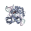
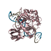
| Structure viewer | Molecule: MolmilJmol/JSmol |
|---|
- Downloads & links
-Download
| PDBx/mmCIF format | 6jxn.cif.gz | 193.1 KB | Display | PDBx/mmCIF format |
|---|---|---|---|---|
| PDB format | pdb6jxn.ent.gz | 151.4 KB | Display | PDB format |
| PDBx/mmJSON format | 6jxn.json.gz | Tree view | PDBx/mmJSON format | |
| Others |  Other downloads Other downloads |
-Validation report
| Arichive directory | https://data.pdbj.org/pub/pdb/validation_reports/jx/6jxnftp://data.pdbj.org/pub/pdb/validation_reports/jx/6jxn | HTTPS FTP |
|---|
-Related structure data
| Related structure data |  6jxsC  3w78S S: Starting model for refinement C: citing same article ( |
|---|---|
| Similar structure data |


-Links
 PDBj
PDBj- Assembly
| Deposited unit | 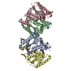
| ||||||||
|---|---|---|---|---|---|---|---|---|---|
| 1 | 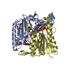
| ||||||||
| 2 | 
| ||||||||
| Unit cell |
|
-Components
| #1: Protein | Mass: 25570.969 Da / Num. of mol.: 4 Source method: isolated from a genetically manipulated source Source: (gene. exp.) References: UniProt: G9QLG5, Oxidoreductases; Acting on other nitrogenous compounds as donors #2: Chemical | ChemComp-FMN /   Mass: 456.344 Da / Num. of mol.: 4 / Source method: obtained synthetically / Formula: C17H21N4O9P / Feature type: SUBJECT OF INVESTIGATION Mass: 456.344 Da / Num. of mol.: 4 / Source method: obtained synthetically / Formula: C17H21N4O9P / Feature type: SUBJECT OF INVESTIGATION#3: Chemical |   Mass: 370.436 Da / Num. of mol.: 2 / Source method: obtained synthetically / Formula: C16H34O9 Mass: 370.436 Da / Num. of mol.: 2 / Source method: obtained synthetically / Formula: C16H34O9#4: Chemical |   Mass: 207.290 Da / Num. of mol.: 3 / Source method: obtained synthetically / Formula: C8H17NO3S / Feature type: SUBJECT OF INVESTIGATION / Comment: pH buffer*YM Mass: 207.290 Da / Num. of mol.: 3 / Source method: obtained synthetically / Formula: C8H17NO3S / Feature type: SUBJECT OF INVESTIGATION / Comment: pH buffer*YM#5: Water | ChemComp-HOH / |  Mass: 18.015 Da / Num. of mol.: 466 / Source method: isolated from a natural source / Formula: H2O Mass: 18.015 Da / Num. of mol.: 466 / Source method: isolated from a natural source / Formula: H2O |
|---|
-Experimental details
-Experiment
| Experiment | Method: X-RAY DIFFRACTION / Number of used crystals: 1 |
|---|
- Sample preparation
| Crystal | Density Matthews: 2.39 Å3/Da / Density % sol: 43.71 % |
|---|---|
| Crystal grow | Temperature: 293 K / Method: vapor diffusion, sitting drop / pH: 9.5 / Details: 40% PEG 600, 0.1 M CHES |
-Data collection
| Diffraction | Mean temperature: 100 K / Serial crystal experiment: N |
|---|---|
| Diffraction source | Source: SYNCHROTRON / Site: Photon Factory  / Beamline: AR-NE3A / Wavelength: 1 Å / Beamline: AR-NE3A / Wavelength: 1 Å |
| Detector | Type: DECTRIS PILATUS 2M / Detector: PIXEL / Date: Nov 8, 2015 |
| Radiation | Protocol: SINGLE WAVELENGTH / Monochromatic (M) / Laue (L): M / Scattering type: x-ray |
| Radiation wavelength | Wavelength: 1 Å / Relative weight: 1 |
| Reflection | Resolution: 1.97→35.62 Å / Num. obs: 62256 / % possible obs: 99.9 % / Redundancy: 5 % / Biso Wilson estimate: 11.2 Å2 / Rmerge(I) obs: 0.159 / Net I/σ(I): 9.8 |
| Reflection shell | Resolution: 1.97→2 Å / Redundancy: 4 % / Rmerge(I) obs: 0.061 / Num. unique obs: 9820 / % possible all: 99.8 |
- Processing
| Software |
| ||||||||||||||||||||
|---|---|---|---|---|---|---|---|---|---|---|---|---|---|---|---|---|---|---|---|---|---|
| Refinement | Method to determine structure: MOLECULAR REPLACEMENT Starting model: 3W78 Resolution: 1.97→35.62 Å / Cross valid method: FREE R-VALUE
| ||||||||||||||||||||
| Displacement parameters | Biso mean: 22 Å2 | ||||||||||||||||||||
| Refinement step | Cycle: LAST / Resolution: 1.97→35.62 Å
| ||||||||||||||||||||
| LS refinement shell | Resolution: 1.97→2 Å /
|
