 Movie
Movie Controller

[English] 日本語
 Yorodumi
- PDB-6ip6: Cryo-EM structure of the CMV-stalled human 80S ribosome with HCV ... -
+ Open data

- Basic information
| Entry | Database: PDB / ID: 6ip6 | ||||||
|---|---|---|---|---|---|---|---|
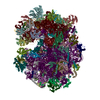
| Title | Cryo-EM structure of the CMV-stalled human 80S ribosome with HCV IRES (Structure iii) | ||||||
 Components Components |
| ||||||
 Keywords Keywords | RIBOSOME / Translation | ||||||
| Function / homology |  Function and homology information Function and homology informationtranslation at presynapse / exit from mitosis / optic nerve development / response to insecticide / regulation of translation involved in cellular response to UV / eukaryotic 80S initiation complex / ribosomal protein import into nucleus / regulation of G1 to G0 transition / negative regulation of endoplasmic reticulum unfolded protein response / axial mesoderm development ...translation at presynapse / exit from mitosis / optic nerve development / response to insecticide / regulation of translation involved in cellular response to UV / eukaryotic 80S initiation complex / ribosomal protein import into nucleus / regulation of G1 to G0 transition / negative regulation of endoplasmic reticulum unfolded protein response / axial mesoderm development / negative regulation of formation of translation preinitiation complex / retinal ganglion cell axon guidance / oxidized pyrimidine DNA binding / response to TNF agonist / positive regulation of base-excision repair / positive regulation of intrinsic apoptotic signaling pathway in response to DNA damage by p53 class mediator / protein-DNA complex disassembly / positive regulation of respiratory burst involved in inflammatory response / positive regulation of gastrulation / 90S preribosome assembly / protein tyrosine kinase inhibitor activity / positive regulation of intrinsic apoptotic signaling pathway in response to DNA damage / positive regulation of ubiquitin-protein transferase activity / IRE1-RACK1-PP2A complex / positive regulation of DNA-templated transcription initiation / positive regulation of Golgi to plasma membrane protein transport / alpha-beta T cell differentiation / nucleolus organization / TNFR1-mediated ceramide production / positive regulation of DNA damage response, signal transduction by p53 class mediator / GAIT complex / negative regulation of RNA splicing / TORC2 complex binding / neural crest cell differentiation / G1 to G0 transition / supercoiled DNA binding / NF-kappaB complex / negative regulation of DNA repair / middle ear morphogenesis / cysteine-type endopeptidase activator activity involved in apoptotic process / oxidized purine DNA binding / cytoplasmic translational initiation / rRNA modification in the nucleus and cytosol / negative regulation of intrinsic apoptotic signaling pathway in response to hydrogen peroxide / regulation of establishment of cell polarity / negative regulation of phagocytosis / negative regulation of bicellular tight junction assembly / ubiquitin-like protein conjugating enzyme binding / erythrocyte homeostasis / cytoplasmic side of rough endoplasmic reticulum membrane / Formation of the ternary complex, and subsequently, the 43S complex / ion channel inhibitor activity / protein kinase A binding / laminin receptor activity / homeostatic process / pigmentation / Ribosomal scanning and start codon recognition / positive regulation of mitochondrial depolarization / Translation initiation complex formation / lung morphogenesis / macrophage chemotaxis / negative regulation of Wnt signaling pathway / positive regulation of natural killer cell proliferation / male meiosis I / fibroblast growth factor binding / Protein hydroxylation / BH3 domain binding / negative regulation of translational frameshifting / regulation of adenylate cyclase-activating G protein-coupled receptor signaling pathway / monocyte chemotaxis / TOR signaling / mTORC1-mediated signalling / SARS-CoV-1 modulates host translation machinery / iron-sulfur cluster binding / positive regulation of GTPase activity / regulation of cell division / cellular response to ethanol / Peptide chain elongation / Selenocysteine synthesis / Formation of a pool of free 40S subunits / negative regulation of protein binding / Eukaryotic Translation Termination / positive regulation of intrinsic apoptotic signaling pathway by p53 class mediator / blastocyst development / protein serine/threonine kinase inhibitor activity / SRP-dependent cotranslational protein targeting to membrane / Response of EIF2AK4 (GCN2) to amino acid deficiency / negative regulation of respiratory burst involved in inflammatory response / ubiquitin ligase inhibitor activity / Viral mRNA Translation / endonucleolytic cleavage to generate mature 3'-end of SSU-rRNA from (SSU-rRNA, 5.8S rRNA, LSU-rRNA) / protein localization to nucleus / negative regulation of ubiquitin-dependent protein catabolic process / Nonsense Mediated Decay (NMD) independent of the Exon Junction Complex (EJC) / positive regulation of signal transduction by p53 class mediator / GTP hydrolysis and joining of the 60S ribosomal subunit / L13a-mediated translational silencing of Ceruloplasmin expression / Major pathway of rRNA processing in the nucleolus and cytosol / protein targeting / regulation of translational fidelity Similarity search - Function | ||||||
| Biological species |  Homo sapiens (human) Homo sapiens (human) | ||||||
| Method | ELECTRON MICROSCOPY / single particle reconstruction / cryo EM / Resolution: 4.5 Å | ||||||
 Authors Authors | Yokoyama, T. / Shigematsu, H. / Shirouzu, M. / Imataka, H. / Ito, T. | ||||||
| Funding support |  Japan, 1items Japan, 1items
| ||||||
 Citation Citation | Journal: Mol Cell / Year: 2019 Title: HCV IRES Captures an Actively Translating 80S Ribosome. Authors: Takeshi Yokoyama / Kodai Machida / Wakana Iwasaki / Tomoaki Shigeta / Madoka Nishimoto / Mari Takahashi / Ayako Sakamoto / Mayumi Yonemochi / Yoshie Harada / Hideki Shigematsu / Mikako ...Authors: Takeshi Yokoyama / Kodai Machida / Wakana Iwasaki / Tomoaki Shigeta / Madoka Nishimoto / Mari Takahashi / Ayako Sakamoto / Mayumi Yonemochi / Yoshie Harada / Hideki Shigematsu / Mikako Shirouzu / Hisashi Tadakuma / Hiroaki Imataka / Takuhiro Ito / Abstract: Translation initiation of hepatitis C virus (HCV) genomic RNA is induced by an internal ribosome entry site (IRES). Our cryoelectron microscopy (cryo-EM) analysis revealed that the HCV IRES binds to ...Translation initiation of hepatitis C virus (HCV) genomic RNA is induced by an internal ribosome entry site (IRES). Our cryoelectron microscopy (cryo-EM) analysis revealed that the HCV IRES binds to the solvent side of the 40S platform of the cap-dependently translating 80S ribosome. Furthermore, we obtained the cryo-EM structures of the HCV IRES capturing the 40S subunit of the IRES-dependently translating 80S ribosome. In the elucidated structures, the HCV IRES "body," consisting of domain III except for subdomain IIIb, binds to the 40S subunit, while the "long arm," consisting of domain II, remains flexible and does not impede the ongoing translation. Biochemical experiments revealed that the cap-dependently translating ribosome becomes a better substrate for the HCV IRES than the free ribosome. Therefore, the HCV IRES is likely to efficiently induce the translation initiation of its downstream mRNA with the captured translating ribosome as soon as the ongoing translation terminates. | ||||||
| History |
|
- Structure visualization
| Movie |
Movie viewer |
|---|---|
| Structure viewer | Molecule: MolmilJmol/JSmol |
- Downloads & links
-Download
| PDBx/mmCIF format | 6ip6.cif.gz | 4.8 MB | Display | PDBx/mmCIF format |
|---|---|---|---|---|
| PDB format | pdb6ip6.ent.gz | Display | PDB format | |
| PDBx/mmJSON format | 6ip6.json.gz | Tree view | PDBx/mmJSON format | |
| Others |  Other downloads Other downloads |
-Validation report
| Arichive directory | https://data.pdbj.org/pub/pdb/validation_reports/ip/6ip6ftp://data.pdbj.org/pub/pdb/validation_reports/ip/6ip6 | HTTPS FTP |
|---|
-Related structure data
| Related structure data | 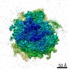 9702MC  9699C 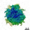 9701C 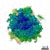 9703C 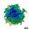 9704C  6ip5C  6ip8C C: citing same article ( M: map data used to model this data |
|---|---|
| Similar structure data |


-Links
 PDBj
PDBj


- Assembly
| Deposited unit | 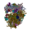
|
|---|---|
| 1 |
|
-Components
-RNA chain , 7 types, 7 molecules 1A1B1C2mzvzyzz
| #1: RNA chain | Mass: 1640182.000 Da / Num. of mol.: 1 / Source method: isolated from a natural source / Source: (natural) Homo sapiens (human) |
|---|---|
| #2: RNA chain | Mass: 38998.078 Da / Num. of mol.: 1 / Source method: isolated from a natural source / Source: (natural) Homo sapiens (human) |
| #3: RNA chain | Mass: 50449.812 Da / Num. of mol.: 1 / Source method: isolated from a natural source / Source: (natural) Homo sapiens (human) |
| #46: RNA chain | Mass: 602752.875 Da / Num. of mol.: 1 / Source method: isolated from a natural source / Source: (natural) Homo sapiens (human) |
| #80: RNA chain | Mass: 1827.141 Da / Num. of mol.: 1 / Source method: isolated from a natural source / Source: (natural) Homo sapiens (human) |
| #82: RNA chain | Mass: 24189.314 Da / Num. of mol.: 1 / Source method: isolated from a natural source / Source: (natural) Homo sapiens (human) |
| #83: RNA chain | Mass: 93491.078 Da / Num. of mol.: 1 / Source method: isolated from a natural source / Source: (natural) Homo sapiens (human) |
+60S ribosomal protein ... , 41 types, 41 molecules 1D1E1F1G1H2A2B2C2D2E2F2G2H2I2J2K2L2M2N2O2P2Q2R2S2T2U2V2W2X2Y...
-Protein , 3 types, 3 molecules 2g3F3R
| #41: Protein | Mass: 14758.394 Da / Num. of mol.: 1 / Source method: isolated from a natural source / Source: (natural) Homo sapiens (human) / References: UniProt: P62987 |
|---|---|
| #67: Protein | Mass: 35115.652 Da / Num. of mol.: 1 / Source method: isolated from a natural source / Source: (natural) Homo sapiens (human) / References: UniProt: P63244 |
| #79: Protein | Mass: 18004.041 Da / Num. of mol.: 1 / Source method: isolated from a natural source / Source: (natural) Homo sapiens (human) / References: UniProt: P62979 |
+40S ribosomal protein ... , 31 types, 31 molecules 2n2o2p2q2r2s2t2u2v2w2x2y2z20213A3B3C3D3E3G3H3I3J3K3L3M3N3O3P3Q
-Protein/peptide , 1 types, 1 molecules zx
| #81: Protein/peptide | Mass: 1865.305 Da / Num. of mol.: 1 / Source method: isolated from a natural source / Source: (natural) Homo sapiens (human) |
|---|
-Details
| Has protein modification | Y |
|---|
-Experimental details
-Experiment
| Experiment | Method: ELECTRON MICROSCOPY |
|---|---|
| EM experiment | Aggregation state: PARTICLE / 3D reconstruction method: single particle reconstruction |
- Sample preparation
| Component | Name: Human 80S ribosome / Type: RIBOSOME / Entity ID: all / Source: NATURAL |
|---|---|
| Source (natural) | Organism: Homo sapiens (human) |
| Buffer solution | pH: 7.5 |
| Specimen | Embedding applied: NO / Shadowing applied: NO / Staining applied: NO / Vitrification applied: YES |
| Specimen support | Grid material: COPPER / Grid mesh size: 300 divisions/in. / Grid type: Quantifoil R1.2/1.3 |
| Vitrification | Instrument: FEI VITROBOT MARK IV / Cryogen name: ETHANE / Humidity: 100 % / Chamber temperature: 277 K |
- Electron microscopy imaging
| Experimental equipment |  Model: Talos Arctica / Image courtesy: FEI Company |
|---|---|
| Microscopy | Model: FEI TECNAI ARCTICA |
| Electron gun | Electron source:  FIELD EMISSION GUN / Accelerating voltage: 200 kV / Illumination mode: FLOOD BEAM FIELD EMISSION GUN / Accelerating voltage: 200 kV / Illumination mode: FLOOD BEAM |
| Electron lens | Mode: BRIGHT FIELD / Nominal magnification: 23500 X / Calibrated magnification: 33557 X / Cs: 2.7 mm / C2 aperture diameter: 50 µm |
| Specimen holder | Cryogen: NITROGEN |
| Image recording | Electron dose: 50 e/Å2 / Detector mode: SUPER-RESOLUTION / Film or detector model: GATAN K2 SUMMIT (4k x 4k) |
| Image scans | Movie frames/image: 40 |
- Processing
| CTF correction | Type: NONE |
|---|---|
| Symmetry | Point symmetry: C1 (asymmetric) |
| 3D reconstruction | Resolution: 4.5 Å / Resolution method: FSC 0.143 CUT-OFF / Num. of particles: 21303 / Symmetry type: POINT |
