 Movie
Movie Controller

[English] 日本語
 Yorodumi
- PDB-6inv: Crystal structure of the CysR-CTLD2 fragment of human MR at acidi... -
+ Open data

- Basic information
| Entry | Database: PDB / ID: 6inv | |||||||||
|---|---|---|---|---|---|---|---|---|---|---|
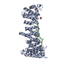
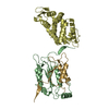
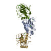
| Title | Crystal structure of the CysR-CTLD2 fragment of human MR at acidic pH (pH 4.0) | |||||||||
 Components Components | Macrophage mannose receptor 1 | |||||||||
 Keywords Keywords | IMMUNE SYSTEM / CD206 / mannose receptor family / pH-dependent / conformational change / scavenger receptor | |||||||||
| Function / homology |  Function and homology information Function and homology informationcargo receptor activity / Cross-presentation of soluble exogenous antigens (endosomes) / D-mannose binding / cellular response to interleukin-4 / receptor-mediated endocytosis / cellular response to type II interferon / Modulation by Mtb of host immune system / transmembrane signaling receptor activity / virus receptor activity / cellular response to lipopolysaccharide ...cargo receptor activity / Cross-presentation of soluble exogenous antigens (endosomes) / D-mannose binding / cellular response to interleukin-4 / receptor-mediated endocytosis / cellular response to type II interferon / Modulation by Mtb of host immune system / transmembrane signaling receptor activity / virus receptor activity / cellular response to lipopolysaccharide / signaling receptor activity / endosome membrane / cell surface / plasma membrane Similarity search - Function | |||||||||
| Biological species |  Homo sapiens (human) Homo sapiens (human) | |||||||||
| Method |  X-RAY DIFFRACTION / SYNCHROTRON / MOLECULAR REPLACEMENT / Resolution: 3.3 Å X-RAY DIFFRACTION / SYNCHROTRON / MOLECULAR REPLACEMENT / Resolution: 3.3 Å | |||||||||
 Authors Authors | Hu, Z. / He, Y. | |||||||||
| Funding support |  China, 2items China, 2items
| |||||||||
 Citation Citation | Journal: J.Struct.Biol. / Year: 2019 Title: Structural basis of the pH-dependent conformational change of the N-terminal region of human mannose receptor/CD206. Authors: Hu, Z. / Wang, Y. / Cheng, C. / He, Y. | |||||||||
| History |
|
- Structure visualization
| Structure viewer | Molecule: MolmilJmol/JSmol |
|---|
- Downloads & links
-Download
| PDBx/mmCIF format | 6inv.cif.gz | 102 KB | Display | PDBx/mmCIF format |
|---|---|---|---|---|
| PDB format | pdb6inv.ent.gz | 75.6 KB | Display | PDB format |
| PDBx/mmJSON format | 6inv.json.gz | Tree view | PDBx/mmJSON format | |
| Others |  Other downloads Other downloads |
-Validation report
| Arichive directory | https://data.pdbj.org/pub/pdb/validation_reports/in/6invftp://data.pdbj.org/pub/pdb/validation_reports/in/6inv | HTTPS FTP |
|---|
-Related structure data
| Related structure data |  6innC  6inoC 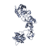 6inuC  6ioeC  5xtsS S: Starting model for refinement C: citing same article ( |
|---|---|
| Similar structure data |


-Links
 PDBj
PDBj


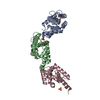
- Assembly
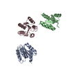
| Deposited unit | 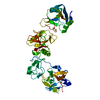
| ||||||||
|---|---|---|---|---|---|---|---|---|---|
| 1 |
| ||||||||
| Unit cell |
|
-Components
| #1: Protein | Mass: 54412.914 Da / Num. of mol.: 1 Source method: isolated from a genetically manipulated source Source: (gene. exp.) Homo sapiens (human) / Gene: MRC1, CLEC13D, CLEC13DL, MRC1L1 / Production host:  Trichoplusia ni (cabbage looper) / References: UniProt: P22897 Trichoplusia ni (cabbage looper) / References: UniProt: P22897 |
|---|---|
| Has protein modification | Y |
-Experimental details
-Experiment
| Experiment | Method: X-RAY DIFFRACTION / Number of used crystals: 1 |
|---|
- Sample preparation
| Crystal | Density Matthews: 2.48 Å3/Da / Density % sol: 50.45 % |
|---|---|
| Crystal grow | Temperature: 291.15 K / Method: vapor diffusion, sitting drop Details: 1.0 M lithium chloride, 0.1 M citric acid (pH 4.0), 10% (w/v) PEG 6000 |
-Data collection
| Diffraction | Mean temperature: 80 K / Serial crystal experiment: N |
|---|---|
| Diffraction source | Source: SYNCHROTRON / Site: SSRF / Beamline: BL18U1 / Wavelength: 0.98 Å |
| Detector | Type: DECTRIS PILATUS3 S 6M / Detector: PIXEL / Date: Dec 2, 2017 |
| Radiation | Protocol: SINGLE WAVELENGTH / Monochromatic (M) / Laue (L): M / Scattering type: x-ray |
| Radiation wavelength | Wavelength: 0.98 Å / Relative weight: 1 |
| Reflection | Resolution: 3.3→50 Å / Num. obs: 8092 / % possible obs: 95.5 % / Redundancy: 6.3 % / Rmerge(I) obs: 0.137 / Net I/σ(I): 2.109 |
| Reflection shell | Resolution: 3.3→3.42 Å / Rmerge(I) obs: 0.283 / Num. unique obs: 738 |
- Processing
| Software |
| |||||||||||||||||||||||||||||||||||||||||||||||||
|---|---|---|---|---|---|---|---|---|---|---|---|---|---|---|---|---|---|---|---|---|---|---|---|---|---|---|---|---|---|---|---|---|---|---|---|---|---|---|---|---|---|---|---|---|---|---|---|---|---|---|
| Refinement | Method to determine structure: MOLECULAR REPLACEMENT Starting model: 5XTS Resolution: 3.3→39.732 Å / SU ML: 0.49 / Cross valid method: FREE R-VALUE / σ(F): 1.4 / Phase error: 30.47
| |||||||||||||||||||||||||||||||||||||||||||||||||
| Solvent computation | Shrinkage radii: 0.9 Å / VDW probe radii: 1.11 Å | |||||||||||||||||||||||||||||||||||||||||||||||||
| Refinement step | Cycle: LAST / Resolution: 3.3→39.732 Å
| |||||||||||||||||||||||||||||||||||||||||||||||||
| Refine LS restraints |
| |||||||||||||||||||||||||||||||||||||||||||||||||
| LS refinement shell |
|
