| Entry | Database: PDB / ID: 6i8n
|
|---|
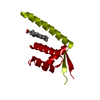
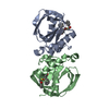
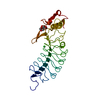
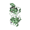
| Title | Crystal structure of LmrR with V15 replaced by unnatural amino acid 4-amino-L-phenylalanine |
|---|
 Components Components | Transcriptional regulator, PadR-like family |
|---|
 Keywords Keywords | DNA BINDING PROTEIN / PadR family / transcriptional regulator / engineered variant / artificial enzyme / unnatural amino acid |
|---|
| Function / homology |  Function and homology information Function and homology information
: / Transcription regulator PadR, N-terminal / Transcriptional regulator PadR-like family / Winged helix-like DNA-binding domain superfamily/Winged helix DNA-binding domain / Arc Repressor Mutant, subunit A / Winged helix DNA-binding domain superfamily / Winged helix-like DNA-binding domain superfamily / Orthogonal Bundle / Mainly AlphaSimilarity search - Domain/homology |
|---|
| Biological species |  Lactococcus lactis subsp. cremoris (lactic acid bacteria) Lactococcus lactis subsp. cremoris (lactic acid bacteria) |
|---|
| Method |  X-RAY DIFFRACTION / SYNCHROTRON / MOLECULAR REPLACEMENT / Resolution: 1.79 Å X-RAY DIFFRACTION / SYNCHROTRON / MOLECULAR REPLACEMENT / Resolution: 1.79 Å |
|---|
 Authors Authors | Reddem, R. / Thunnissen, A.M.W.H. |
|---|
| Funding support |  Netherlands, 1items Netherlands, 1items | Organization | Grant number | Country |
|---|
| Netherlands Organisation for Scientific Research | 724.013.003 | Netherlands |
|
|---|
 Citation Citation | Journal: Angew. Chem. Int. Ed. Engl. / Year: 2019
Title: Directed Evolution of a Designer Enzyme Featuring an Unnatural Catalytic Amino Acid.
Authors: Mayer, C. / Dulson, C. / Reddem, E. / Thunnissen, A.W.H. / Roelfes, G. |
|---|
| History | | Deposition | Nov 20, 2018 | Deposition site: PDBE / Processing site: PDBE |
|---|
| Revision 1.0 | Jan 2, 2019 | Provider: repository / Type: Initial release |
|---|
| Revision 1.1 | Feb 13, 2019 | Group: Data collection / Database references / Category: citation / citation_author / pdbx_database_proc
Item: _citation.journal_volume / _citation.page_first ..._citation.journal_volume / _citation.page_first / _citation.page_last / _citation.title / _citation.year / _citation_author.identifier_ORCID / _citation_author.name |
|---|
| Revision 1.2 | Jan 24, 2024 | Group: Data collection / Database references / Refinement description
Category: chem_comp_atom / chem_comp_bond ...chem_comp_atom / chem_comp_bond / database_2 / pdbx_initial_refinement_model / struct_ncs_dom_lim
Item: _database_2.pdbx_DOI / _database_2.pdbx_database_accession ..._database_2.pdbx_DOI / _database_2.pdbx_database_accession / _struct_ncs_dom_lim.beg_auth_comp_id / _struct_ncs_dom_lim.beg_label_asym_id / _struct_ncs_dom_lim.beg_label_comp_id / _struct_ncs_dom_lim.beg_label_seq_id / _struct_ncs_dom_lim.end_auth_comp_id / _struct_ncs_dom_lim.end_label_asym_id / _struct_ncs_dom_lim.end_label_comp_id / _struct_ncs_dom_lim.end_label_seq_id |
|---|
|
|---|
 Movie
Movie Controller

 Yorodumi
 Open data
 Basic information
 Structure visualization
 Downloads & links
 Other downloads
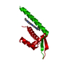


 PDBj
PDBj Assembly