+ Open data
- Basic information
| Entry | Database: PDB / ID: 6fuu | ||||||
|---|---|---|---|---|---|---|---|
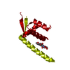
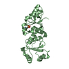
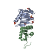
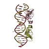
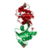
| Title | Transcriptional regulator LmrR with bound heme | ||||||
 Components Components | Transcriptional regulator PadR family | ||||||
 Keywords Keywords | DNA BINDING PROTEIN / Artificial enzyme / Complex / Heme-based catalysis / Transcriptional regulator / Multi-drug resistance | ||||||
| Function / homology |  Function and homology information Function and homology informationTranscription regulator PadR, N-terminal / Transcriptional regulator PadR-like family / Winged helix-like DNA-binding domain superfamily/Winged helix DNA-binding domain / Arc Repressor Mutant, subunit A / Winged helix DNA-binding domain superfamily / Winged helix-like DNA-binding domain superfamily / Orthogonal Bundle / Mainly Alpha Similarity search - Domain/homology | ||||||
| Biological species |  Lactococcus lactis subsp. cremoris (lactic acid bacteria) Lactococcus lactis subsp. cremoris (lactic acid bacteria) | ||||||
| Method |  X-RAY DIFFRACTION / X-RAY DIFFRACTION /  SYNCHROTRON / SYNCHROTRON /  MOLECULAR REPLACEMENT / Resolution: 1.75 Å MOLECULAR REPLACEMENT / Resolution: 1.75 Å | ||||||
 Authors Authors | Reddem, E.R. / Thunnissen, A.M.W.H. | ||||||
| Funding support |  Netherlands, 1items Netherlands, 1items
| ||||||
 Citation Citation |  Journal: Angew. Chem. Int. Ed. Engl. / Year: 2018 Journal: Angew. Chem. Int. Ed. Engl. / Year: 2018Title: An Artificial Heme Enzyme for Cyclopropanation Reactions. Authors: Villarino, L. / Splan, K.E. / Reddem, E. / Alonso-Cotchico, L. / Gutierrez de Souza, C. / Lledos, A. / Marechal, J.D. / Thunnissen, A.W.H. / Roelfes, G. | ||||||
| History |
|
- Structure visualization
| Structure viewer | Molecule:  Molmil Molmil Jmol/JSmol Jmol/JSmol |
|---|
- Downloads & links
- Download
| PDBx/mmCIF format |  6fuu.cif.gz 6fuu.cif.gz | 59.6 KB | Display |  PDBx/mmCIF format PDBx/mmCIF format |
|---|---|---|---|---|
| PDB format |  pdb6fuu.ent.gz pdb6fuu.ent.gz | 43.4 KB | Display |  PDB format PDB format |
| PDBx/mmJSON format |  6fuu.json.gz 6fuu.json.gz | Tree view |  PDBx/mmJSON format PDBx/mmJSON format | |
| Others |  Other downloads Other downloads |
-Validation report
| Arichive directory |  https://data.pdbj.org/pub/pdb/validation_reports/fu/6fuu https://data.pdbj.org/pub/pdb/validation_reports/fu/6fuu ftp://data.pdbj.org/pub/pdb/validation_reports/fu/6fuu ftp://data.pdbj.org/pub/pdb/validation_reports/fu/6fuu | HTTPS FTP |
|---|
-Related structure data
| Related structure data | 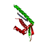 3f8cS S: Starting model for refinement |
|---|---|
| Similar structure data |
- Links
Links
- Assembly
| Deposited unit | 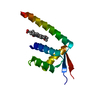
| ||||||||||||
|---|---|---|---|---|---|---|---|---|---|---|---|---|---|
| 1 | 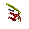
| ||||||||||||
| Unit cell |
| ||||||||||||
| Components on special symmetry positions |
|
- Components
| #1: Protein | Mass: 14764.789 Da / Num. of mol.: 1 Source method: isolated from a genetically manipulated source Details: Residues 1-4, 70-73 and 109-126 are not included in the model due to weak/absent electron density Source: (gene. exp.)  Lactococcus lactis subsp. cremoris (lactic acid bacteria) Lactococcus lactis subsp. cremoris (lactic acid bacteria)Gene: NCDO763_1045, VN96_2738 / Production host:  |
|---|---|
| #2: Chemical | ChemComp-HEM / |
| #3: Water | ChemComp-HOH / |
-Experimental details
-Experiment
| Experiment | Method:  X-RAY DIFFRACTION / Number of used crystals: 1 X-RAY DIFFRACTION / Number of used crystals: 1 |
|---|
- Sample preparation
| Crystal | Density Matthews: 1.89 Å3/Da / Density % sol: 34.9 % |
|---|---|
| Crystal grow | Temperature: 291 K / Method: vapor diffusion, hanging drop / pH: 6.5 Details: 100 mM cacodylate, 300 mM sodium acetate, 25% PEG 200 MME, 200 uM hemin |
-Data collection
| Diffraction | Mean temperature: 100 K | ||||||||||||||||||||||||||||||
|---|---|---|---|---|---|---|---|---|---|---|---|---|---|---|---|---|---|---|---|---|---|---|---|---|---|---|---|---|---|---|---|
| Diffraction source | Source:  SYNCHROTRON / Site: SYNCHROTRON / Site:  ESRF ESRF  / Beamline: MASSIF-3 / Wavelength: 0.9677 Å / Beamline: MASSIF-3 / Wavelength: 0.9677 Å | ||||||||||||||||||||||||||||||
| Detector | Type: DECTRIS EIGER X 4M / Detector: PIXEL / Date: May 21, 2017 | ||||||||||||||||||||||||||||||
| Radiation | Protocol: SINGLE WAVELENGTH / Monochromatic (M) / Laue (L): M / Scattering type: x-ray | ||||||||||||||||||||||||||||||
| Radiation wavelength | Wavelength: 0.9677 Å / Relative weight: 1 | ||||||||||||||||||||||||||||||
| Reflection | Resolution: 1.75→45.17 Å / Num. obs: 12313 / % possible obs: 99.6 % / Redundancy: 9.1 % / Biso Wilson estimate: 29.87 Å2 / CC1/2: 0.997 / Rmerge(I) obs: 0.074 / Rpim(I) all: 0.025 / Rrim(I) all: 0.078 / Net I/σ(I): 16.3 / Num. measured all: 112667 / Scaling rejects: 1 | ||||||||||||||||||||||||||||||
| Reflection shell | Diffraction-ID: 1
|
- Processing
| Software |
| ||||||||||||||||||||||||||||||||||||||||
|---|---|---|---|---|---|---|---|---|---|---|---|---|---|---|---|---|---|---|---|---|---|---|---|---|---|---|---|---|---|---|---|---|---|---|---|---|---|---|---|---|---|
| Refinement | Method to determine structure:  MOLECULAR REPLACEMENT MOLECULAR REPLACEMENTStarting model: PDB entry 3F8C Resolution: 1.75→34.498 Å / SU ML: 0.25 / Cross valid method: THROUGHOUT / σ(F): 1.36 / Phase error: 27.88 / Stereochemistry target values: ML
| ||||||||||||||||||||||||||||||||||||||||
| Solvent computation | Shrinkage radii: 0.9 Å / VDW probe radii: 1.11 Å / Solvent model: FLAT BULK SOLVENT MODEL | ||||||||||||||||||||||||||||||||||||||||
| Displacement parameters | Biso max: 92.13 Å2 / Biso mean: 39.2471 Å2 / Biso min: 17.09 Å2 | ||||||||||||||||||||||||||||||||||||||||
| Refinement step | Cycle: final / Resolution: 1.75→34.498 Å
| ||||||||||||||||||||||||||||||||||||||||
| Refine LS restraints |
| ||||||||||||||||||||||||||||||||||||||||
| LS refinement shell | Refine-ID: X-RAY DIFFRACTION / Rfactor Rfree error: 0 / Total num. of bins used: 4
| ||||||||||||||||||||||||||||||||||||||||
| Refinement TLS params. | Method: refined / Origin x: 20.9142 Å / Origin y: 18.6764 Å / Origin z: 123.0833 Å
| ||||||||||||||||||||||||||||||||||||||||
| Refinement TLS group | Selection details: chain 'A' and (resid 5 through 108 ) |
 Movie
Movie Controller





 PDBj
PDBj


